Erik Taylor1
1Radiology, University of New Mexico, Albuquerque, NM, United States
1Radiology, University of New Mexico, Albuquerque, NM, United States
The purpose of this study was to relate brain pathology that develops with advanced age to vascular parameters in normal and hypertensive rodents. Brain pathology was detected by multiple mechanisms of MRI contrast, while specific vascular parameters were derived from ultrasound in the aorta.
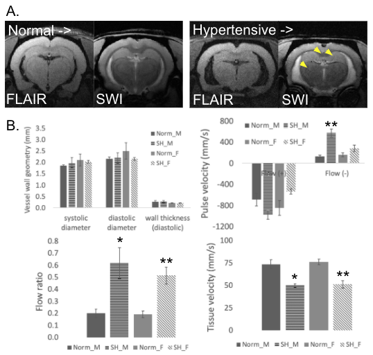
Figure 1. MRI and ultrasound in aged normal (WKY) and hypertensive rats (SHR). A) The brains of aged (14-19 mo) WKY and SHR were characterized by multiple MRI contrast mechanisms to detect lesions. SHR had enlarged ventricles and more CMBs (yellow arrow heads) when compared to WKY. B) Quantification of ultrasound parameters in the same aged animals was performed including vessel wall structure, pulse velocity, flow ratio (calculated), and tissue velocity. The * indicates a P value less than 0.05 and the ** indicates a P value less than 0.01.