Xiaole Zhong1 and J. Jean Chen1,2
1Rotman Research Institute, Baycrest Health Sciences, Toronto, ON, Canada, 2Department of Medical Biophysics, University of Toronto, Toronto, ON, Canada
1Rotman Research Institute, Baycrest Health Sciences, Toronto, ON, Canada, 2Department of Medical Biophysics, University of Toronto, Toronto, ON, Canada
The frequency and amplitude of EEG bands both changed with age, but there is no association between these changes and corresponding parameters in the resting-state fMRI signal. The ratio of fMRI to EEG power is reduced in older adults.
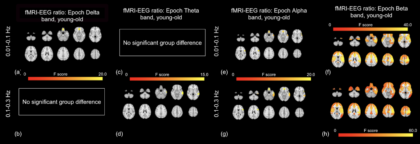
Figure 2. rs-fMRI-EEG power ratio versus age: epoch-based EEG. The fMRI-to-beta power ratio (SDratio) exhibits the greatest age effects (young > old) (f, h). The effects are most pronounced in the 0.1-0.3 Hz range, and the affected regions span nearly the entire anterior cortex, also including subcortical regions such as the putamen and pallidum. The fMRI-to-theta ratio is lower in the young subjects in only the paracentral cortex.
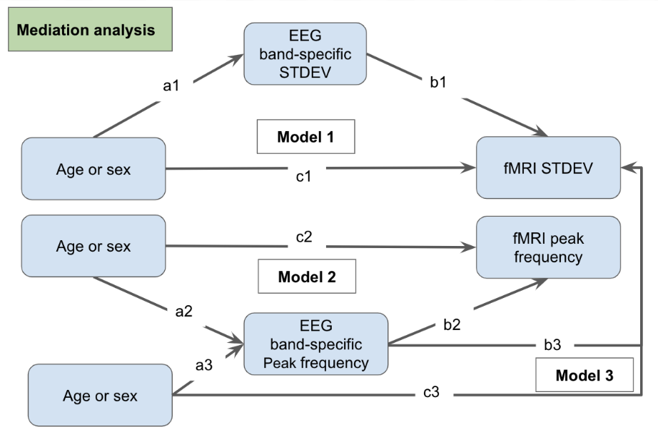
Figure 1. Models for the mediation analyses