Caitlin F Fowler1,2, Dan Madularu3, Gabriel A Devenyi4,5, John Breitner5,6, and Jamie Near1,4,5
1Biological and Biomedical Engineering, McGill University, Montreal, QC, Canada, 2Cerebral Imaging Centre, Douglas Hospital Research Centre, Verdun, QC, Canada, 3Center for Translational Neuroimaging, Northeastern University, Boston, MA, United States, 4Cerebral Imaging Centre, Douglas Hospital Research Institute, Verdun, QC, Canada, 5Psychiatry, McGill University, Montreal, QC, Canada, 6Division of Human Neurosciences, Douglas Hospital Research Centre, Verdun, QC, Canada
1Biological and Biomedical Engineering, McGill University, Montreal, QC, Canada, 2Cerebral Imaging Centre, Douglas Hospital Research Centre, Verdun, QC, Canada, 3Center for Translational Neuroimaging, Northeastern University, Boston, MA, United States, 4Cerebral Imaging Centre, Douglas Hospital Research Institute, Verdun, QC, Canada, 5Psychiatry, McGill University, Montreal, QC, Canada, 6Division of Human Neurosciences, Douglas Hospital Research Centre, Verdun, QC, Canada
Our longitudinal neuroimaging study demonstrates the TgF344-AD rat recapitulates most neurochemical features of human AD and that early treatment with Naproxen is more effective than late treatment at mitigating disease-related neurochemical changes.
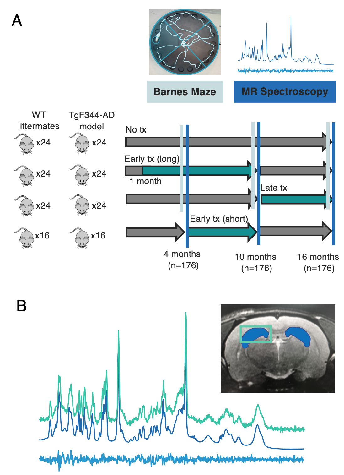
Figure 1: Visualization of study paradigm and spectroscopy data quality. A. MRS scans and Barnes Maze testing were performed in TgF344-AD and WT littermates at 4, 10, and 16 months. Rats were randomly separated into three groups treated with Naproxen: early long treatment, early short treatment, and late treatment. B. Localized 1H-MRS spectra from the hippocampus of a 4-month-old TgF344-AD rat acquired using the PRESS pulse sequence at 7T. Voxel placement is shown in the top right.
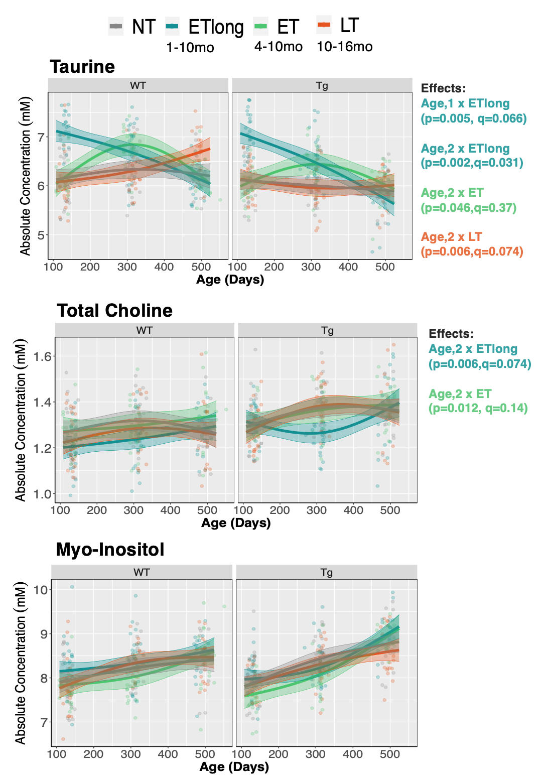
Figure 3: Early but not late treatment mitigates some disease-related metabolic changes. Three treatment paradigms were tested, with timing of Naproxen administration denoted in the figure legend. Linear mixed effects modelling was applied to examine age*genotype*treatment effects. Each rat is depicted by an individual data point. The linear model used to fit the data is represented by a line of best fit and 95% prediction interval (shaded). FDR correction applied at 5%.