M. Dylan Tisdall1, Daniel Ohm2, Rebecca Lobrovich2, Sandhitsu R Das1, Gabor Mizsei1, Karthik Prabhakaran2, Ranjit Ittyerah1, Sydney Lim1, Corey T McMillan2, James Gee1, John Q Trojanowski3, Edward B Lee3, David Wolk2, John A Detre1,2, Paul Yushkevich1, Murray Grossman2, and David Irwin2
1Radiology, Perelman School of Medicine, University of Pennsylvania, Philadelphia, PA, United States, 2Neurology, Perelman School of Medicine, University of Pennsylvania, Philadelphia, PA, United States, 3Pathology and Laboratory Medicine, Perelman School of Medicine, University of Pennsylvania, Philadelphia, PA, United States
1Radiology, Perelman School of Medicine, University of Pennsylvania, Philadelphia, PA, United States, 2Neurology, Perelman School of Medicine, University of Pennsylvania, Philadelphia, PA, United States, 3Pathology and Laboratory Medicine, Perelman School of Medicine, University of Pennsylvania, Philadelphia, PA, United States
We demonstrate that T2*-weighted ex vivo imaging of brain hemispheres from patients with frontotemporal lobar degeneration detects focal iron-rich pathology within the cortex and adjacent WM, validated via MRI-guided histopathology.
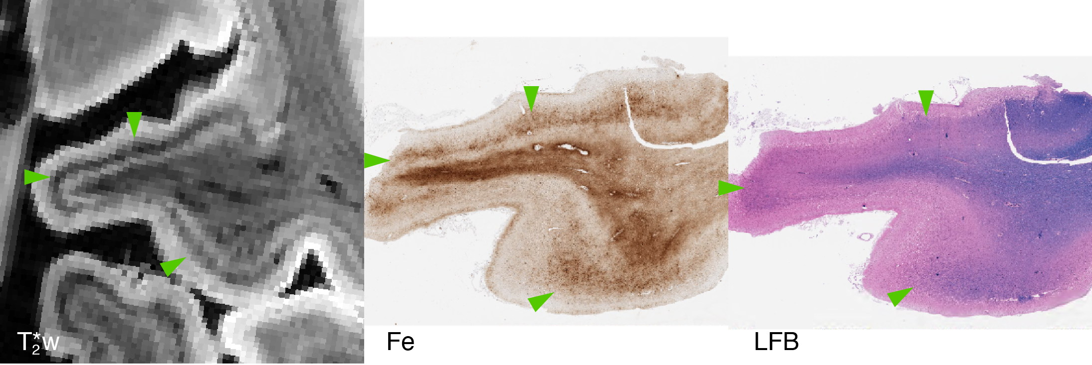
Fig 2. Sample from motor cortex in FTLD-tau with four-repeat GGT tauopathy and clinical naPPA. Note the dark band in the middle layers of cortex, progressing along the entirety of the gyrus (green arrows) in the T2*w image (left). This pattern correlates with a similar band of iron (middle). We note that this band overlaps, but is not well-correlated with, myelinated radial fibers on LFB, which show minimal degeneration despite the high degree of gliosis (right).
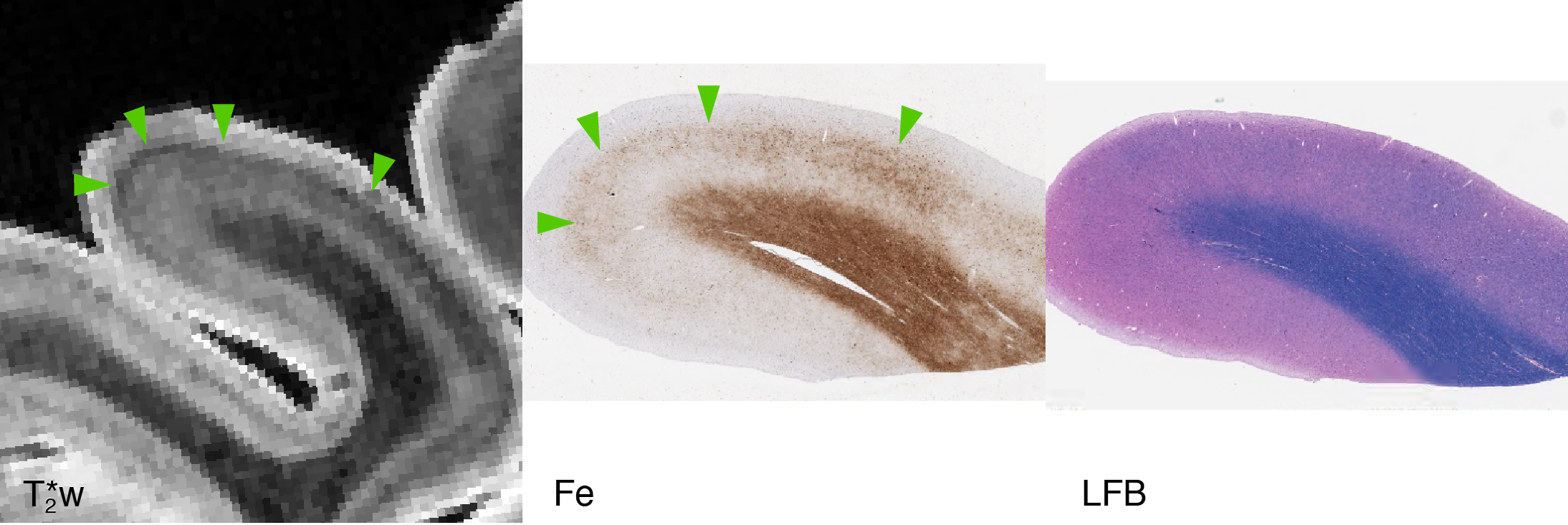
Fig 1. Sample from motor cortex in FTLD-tau with four-repeat tauopathy and clinical PSP. Note the dark band in the middle layers of cortex, broadening as it progresses down the right side of the gyrus (green arrows) in the T2*w image (left). This pattern correlates with a similar band of iron (middle), but not with a particular change in tissue structure or myelination on LFB (right).