Susana Muñoz Maniega1, Jonathan D Clayden2, Maria Valdés Hernandez1, Mark E Bastin1, Ian J Deary3, and Joanna M Wardlaw1
1Centre for Clinical Brain Sciences, University of Edinburgh, Edinburgh, United Kingdom, 2UCL GOS Institute of Child Health, University College London, London, United Kingdom, 3Department of Psychology, University of Edinburgh, Edinburgh, United Kingdom
1Centre for Clinical Brain Sciences, University of Edinburgh, Edinburgh, United Kingdom, 2UCL GOS Institute of Child Health, University College London, London, United Kingdom, 3Department of Psychology, University of Edinburgh, Edinburgh, United Kingdom
Automated methods produced reasonable tract segmentations in healthy older brains. However, both visual assessments and the estimated overlaps between segmented bundles suggest there is a key trade-off between tract coverage and the specificity of associated diffusion parameters.
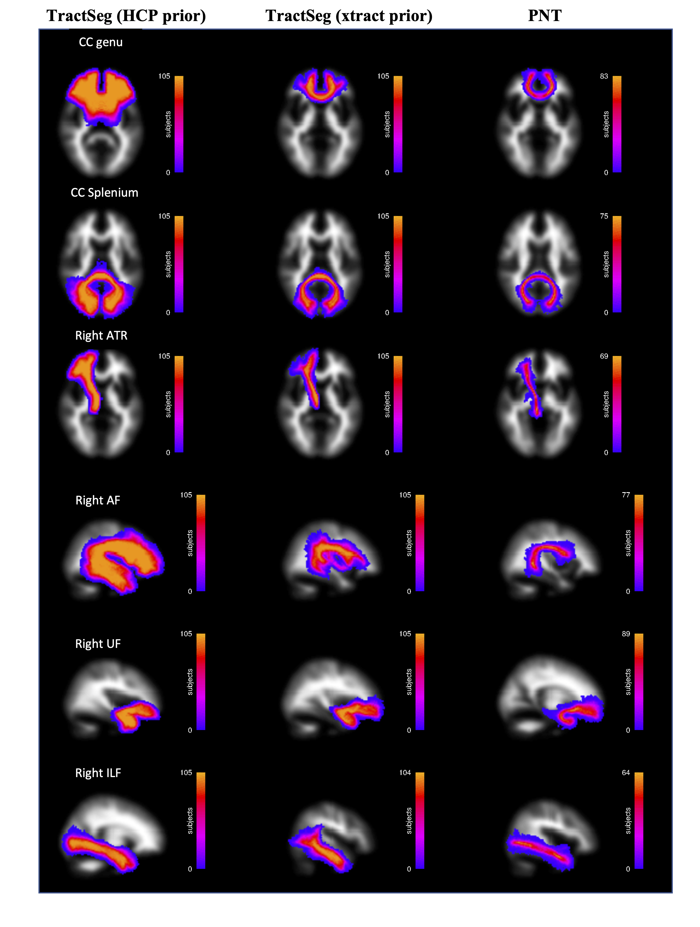
Figure 1. Group maps of white matter tracts segmented with three methods. Colour scales show the voxel-wise frequency for each tract (total N=105).
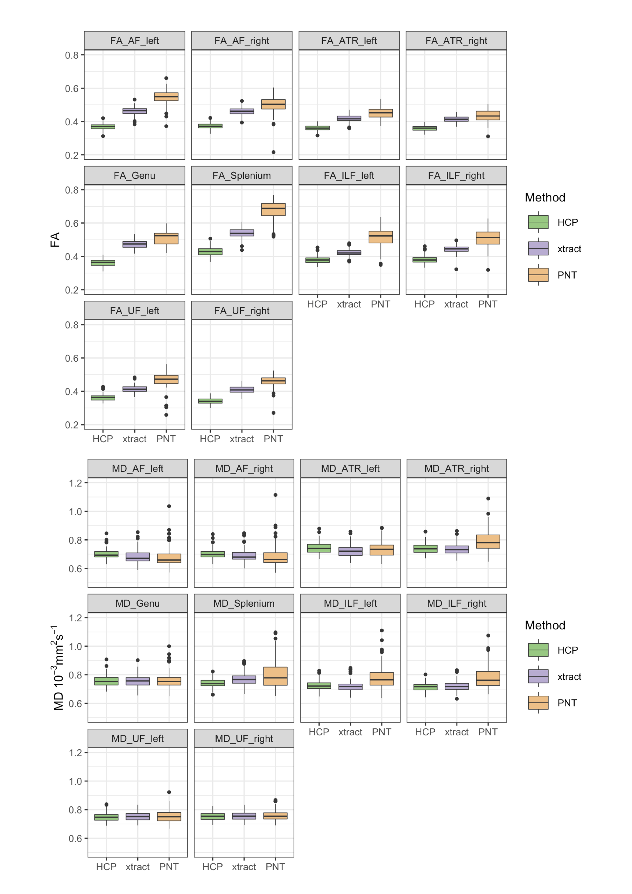
Figure 2. Boxplots of FA and MD obtained in each segmented tract using each method.