Adam S Bernstein1, Steve Z Rapcsak2, Michael Hornberger3, and Manojkumar Saranathan4
1College of Medicine, University of Arizona, Tucson, AZ, United States, 2Department of Neurology, University of Arizona, Tucson, AZ, United States, 3Department of Medicine, University of East Anglia, Norwich, United Kingdom, 4Department of Medical Imaging, University of Arizona, Tucson, AZ, United States
1College of Medicine, University of Arizona, Tucson, AZ, United States, 2Department of Neurology, University of Arizona, Tucson, AZ, United States, 3Department of Medicine, University of East Anglia, Norwich, United Kingdom, 4Department of Medical Imaging, University of Arizona, Tucson, AZ, United States
TBD
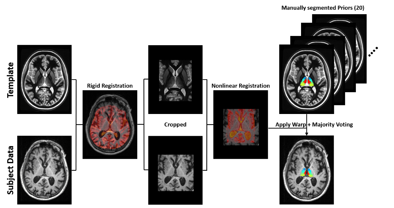
Figure 1. Multi-atlas segmentation scheme for thalamic nuclei segmentation. The multi-atlas consists of 20 manually segmented WMn MPRAGE data which are warped to subject space and label fused using a majority voting scheme. A WMn template is used as an intermediate step to improve robustness and cropping is performed to improve speed and accuracy.
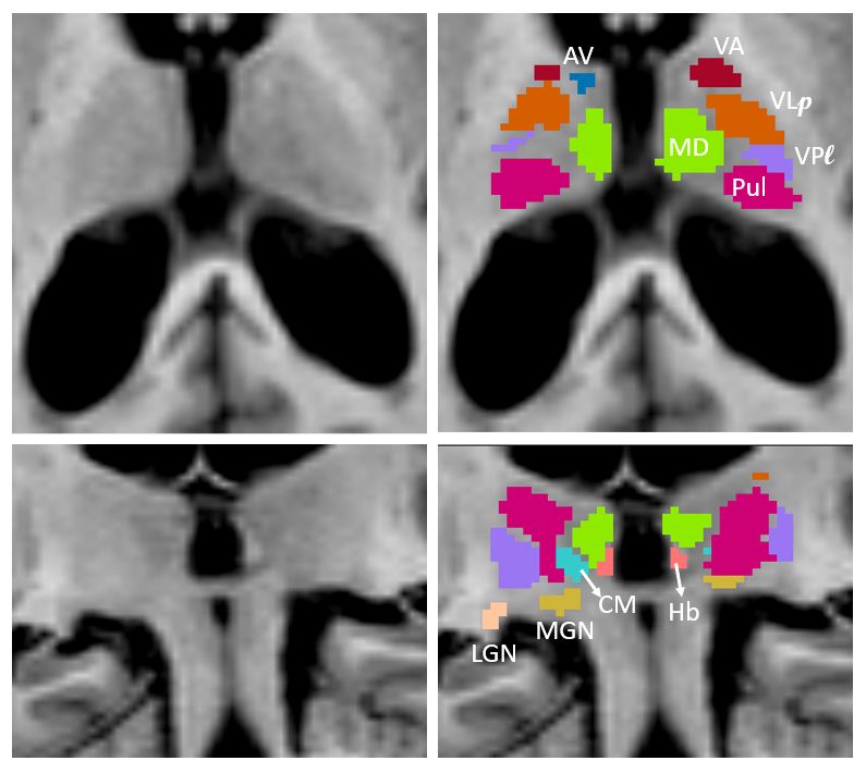
Figure 2. Thalamic nuclei segmentation labels from the modified THOMAS method
overlaid on MPRAGE data in axial (top panel) and coronal (bottom panel) for a patient with AD (enlarged ventricles). Note visualization of small structures such as anteroventral (AV), centromedian (CM) and habenula (Hb).