Brittany Intzandt1,2,3, Safa Sanami4, Julia Huck4, Richard D Hoge5, Louis Bherer2,3,6,7, and Claudine J Gauthier3,4,6
1INDI Department, Concordia University, Montreal, QC, Canada, 2Centre de Recherche de l'Institut Universitaire de Geriatrie, Montreal, QC, Canada, 3Centre de Recherche, l'Institut de Cardiologie de Montréal, Montreal, QC, Canada, 4Physics Department, Concordia University, Montreal, QC, Canada, 5Department of Neurology and Neurosurgery, McGill University, Montreal, QC, Canada, 6PERFORM Centre, Concordia Univeristy, Montreal, QC, Canada, 7Départment de Médicine, Université de Montréal, Montreal, QC, Canada
1INDI Department, Concordia University, Montreal, QC, Canada, 2Centre de Recherche de l'Institut Universitaire de Geriatrie, Montreal, QC, Canada, 3Centre de Recherche, l'Institut de Cardiologie de Montréal, Montreal, QC, Canada, 4Physics Department, Concordia University, Montreal, QC, Canada, 5Department of Neurology and Neurosurgery, McGill University, Montreal, QC, Canada, 6PERFORM Centre, Concordia Univeristy, Montreal, QC, Canada, 7Départment de Médicine, Université de Montréal, Montreal, QC, Canada
Cardiovascular
fitness did not moderate the relationship between increased adiposity and cortical
thickness in overweight males and females, nor in those with normal weight.
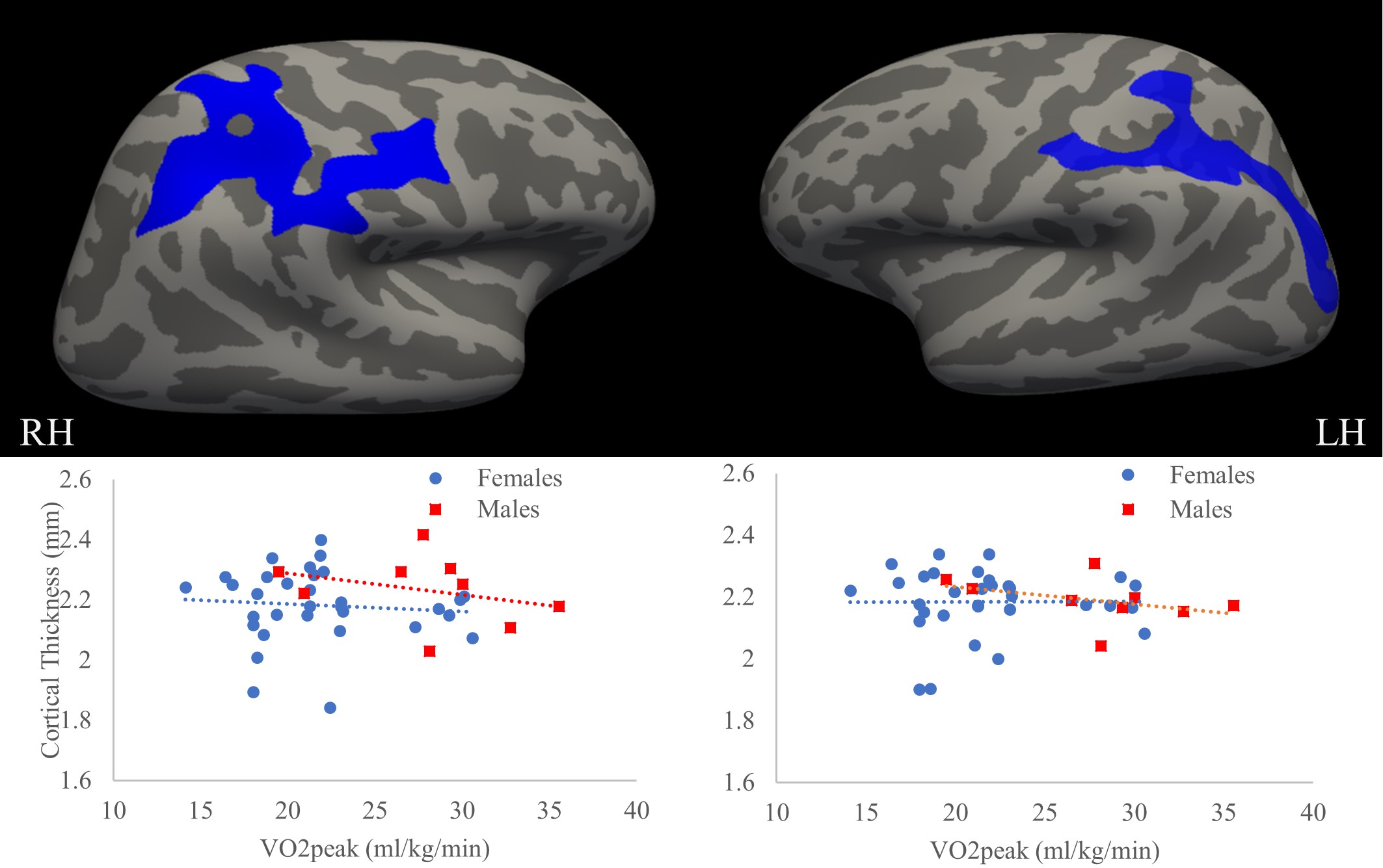
Figure 2: Regions in the right and left hemispheres
(RH; LH) demonstrating significant differences in OBW versus OBM, where OBW demonstrated
greater cortical thickness in these regions indicated in blue (p = 0.00010,
corrected for age, education, study). The graphs demonstrate no influence of
VO2peak on cortical fitness and the relationships for OBW and OBM separately in
the RH (graph on the left; p > 0.05) and LH (graph on the right; p >
0.05).
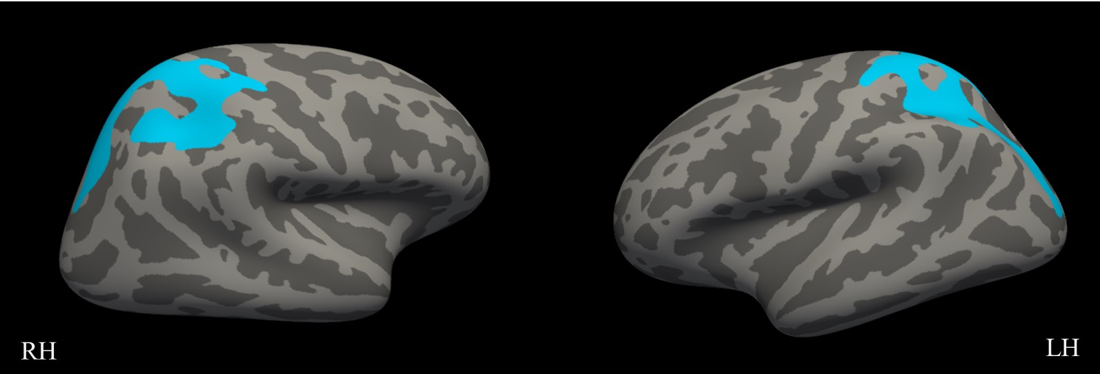
Figure 1: Regions in the right and left hemispheres
(RH; LH) demonstrating significant differences in females versus males, where
females demonstrated greater cortical thickness in these regions in teal
(regardless of BMI status). (p =0.00010; corrected for age, education, study)