Zilin Chen1, Jianpan Huang1, Yang Liu1, Joseph H.C. Lai1, and Kannie W.Y. Chan1,2,3
1Biomedical Engineering, City University of Hong Kong, Hong Kong, Hong Kong, 2Russell H. Morgan Department of Radiology and Radiological Science, The Johns Hopkins University School of Medicine, Baltimore, MD, United States, 3City University of Hong Kong Shenzhen Research Institute, Shenzhen, China
1Biomedical Engineering, City University of Hong Kong, Hong Kong, Hong Kong, 2Russell H. Morgan Department of Radiology and Radiological Science, The Johns Hopkins University School of Medicine, Baltimore, MD, United States, 3City University of Hong Kong Shenzhen Research Institute, Shenzhen, China
Enhanced D-glucose clearance in CSF was observed in AD mice after IL-33 treatment through DGE-MRI at 3T, which could provide a non-invasive way to evaluate AD treatment that facilitate brain lymphatic clearance.
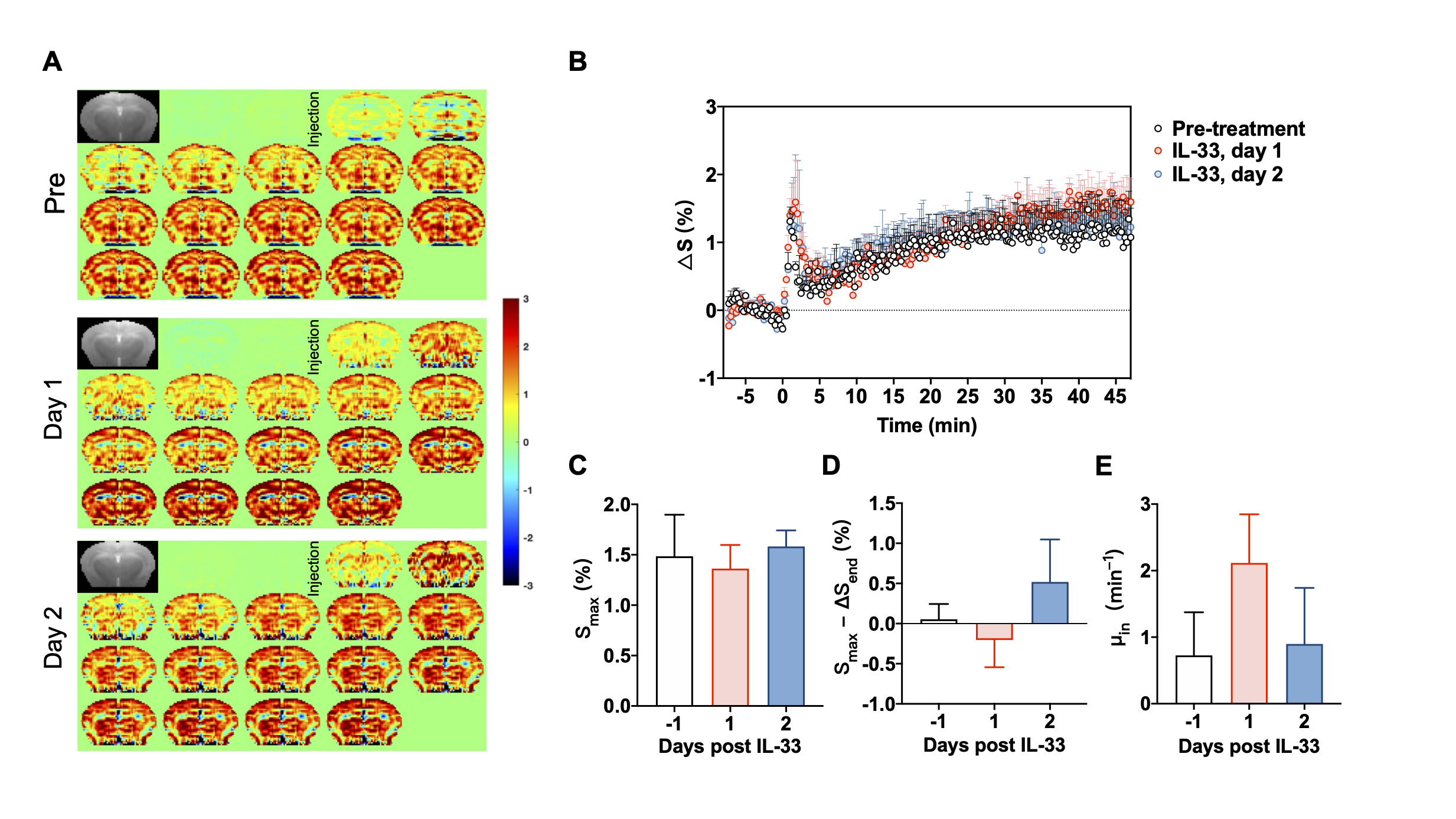
DGE-MRI results for brain parenchyma in APP/PS1 mice before and after IL-33 treatment. (A) Dynamic difference images in brain parenchyma. (B) Experimental DGE plots for brain parenchyma. (C-E) Comparison of the maximum signal Smax, clearance parameters Smax-△Send and glucose uptake rate μin before and after treatment. Data were presented as mean ± SEM (n=4).
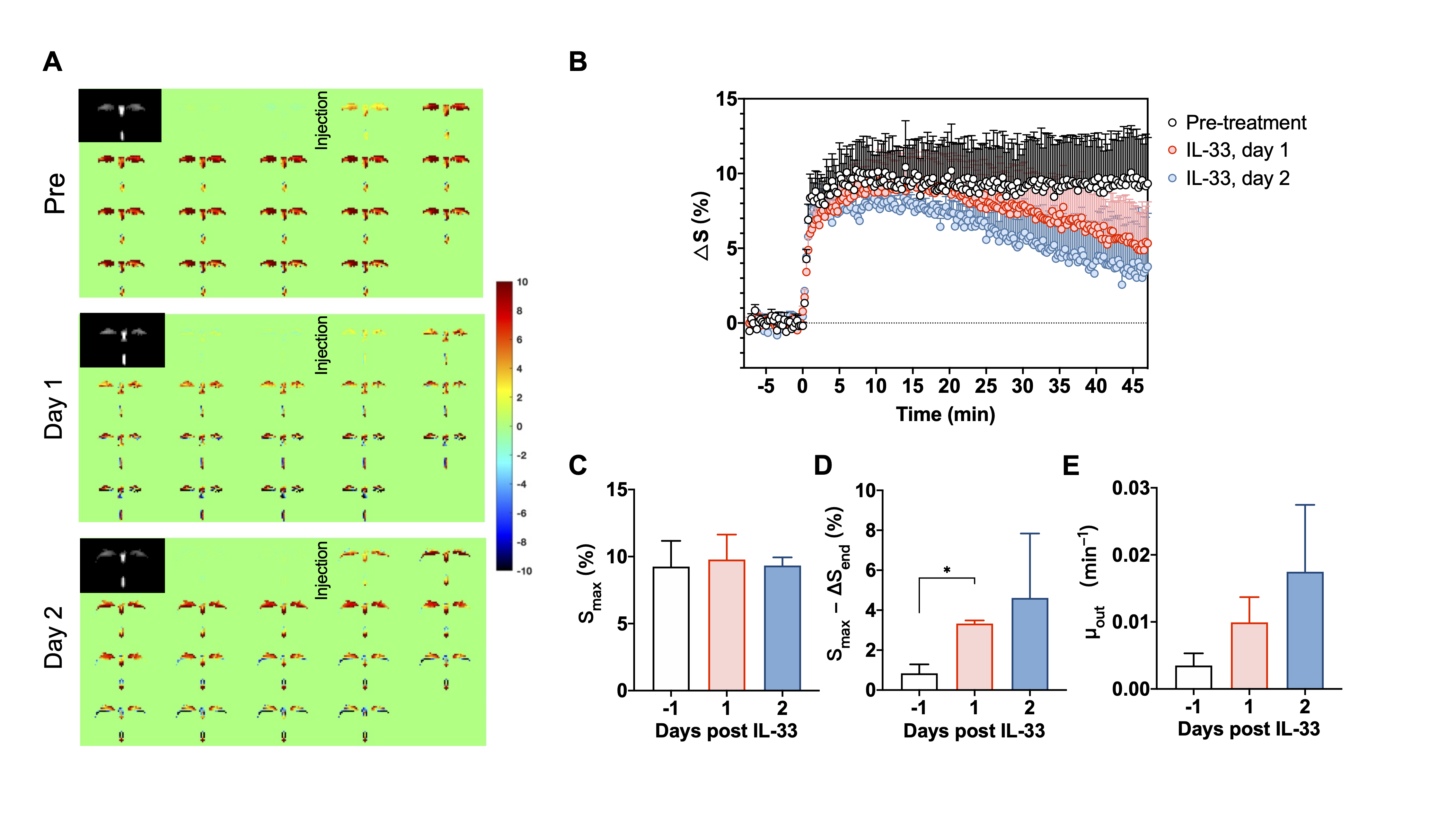
DGE-MRI results for CSF in APP/PS1 mice before and after IL-33 treatment. (A) Dynamic difference images in CSF. (B) Experimental DGE plots for CSF in APP/PS1 mice. (C-E) Comparison of the maximum signal Smax, clearance parameters Smax-△Send and washout rate μout before and after treatment. Data were presented as mean ± SEM (n=4). *P<0.05, one-way ANOVA.