Brenton James Keller1, Jorge Jovicich2, Himanshu Joshi3, Leon Aksman4, John John3, A. B. Dey5, Arthur Toga4, Eileen Crimmins6, and Jinkook Lee1
1CSCR, University of Southern California, Los Angeles, CA, United States, 2Center for Mind/Brain Sciences (CIMEC),, University of Trento, Trento, Italy, 3Multimodal Brain Image Analysis Laboratory, National Institute of Mental Health and Neurosciences, Bangalore, India, 4Laboratory of Neuro Imaging, University of Southern California, Los Angeles, CA, United States, 5All India Institute of Medical Sciences, New Delhi, India, 6Davis School of Gerontology, University of Southern California, Los Angeles, CA, United States
1CSCR, University of Southern California, Los Angeles, CA, United States, 2Center for Mind/Brain Sciences (CIMEC),, University of Trento, Trento, Italy, 3Multimodal Brain Image Analysis Laboratory, National Institute of Mental Health and Neurosciences, Bangalore, India, 4Laboratory of Neuro Imaging, University of Southern California, Los Angeles, CA, United States, 5All India Institute of Medical Sciences, New Delhi, India, 6Davis School of Gerontology, University of Southern California, Los Angeles, CA, United States
Using multicentric brain structural 3T MRI morphometry, we
show evidence in support of the cognitive reserve hypothesis in an Indian
population including healthy elderly (55) and mild cognitively impaired (75)
volunteers.
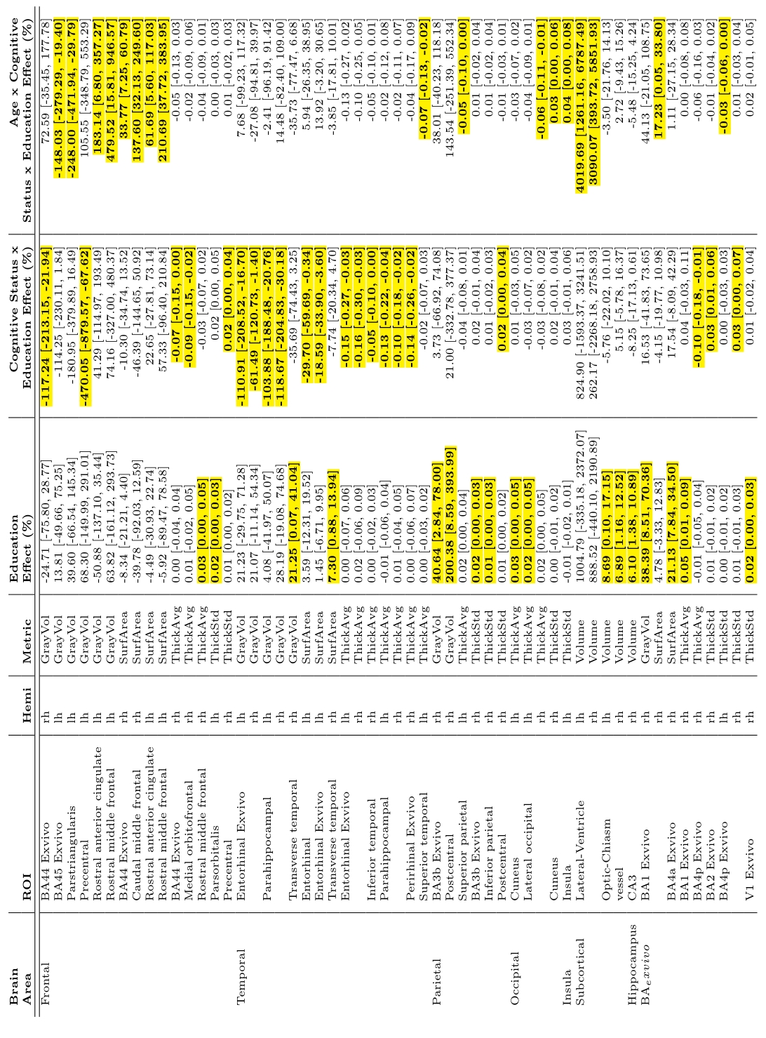
List of brain regions and morphometry metrics that were
found to be significantly associated (highlighted) with either education,
education x disease status, or age x education x disease status.
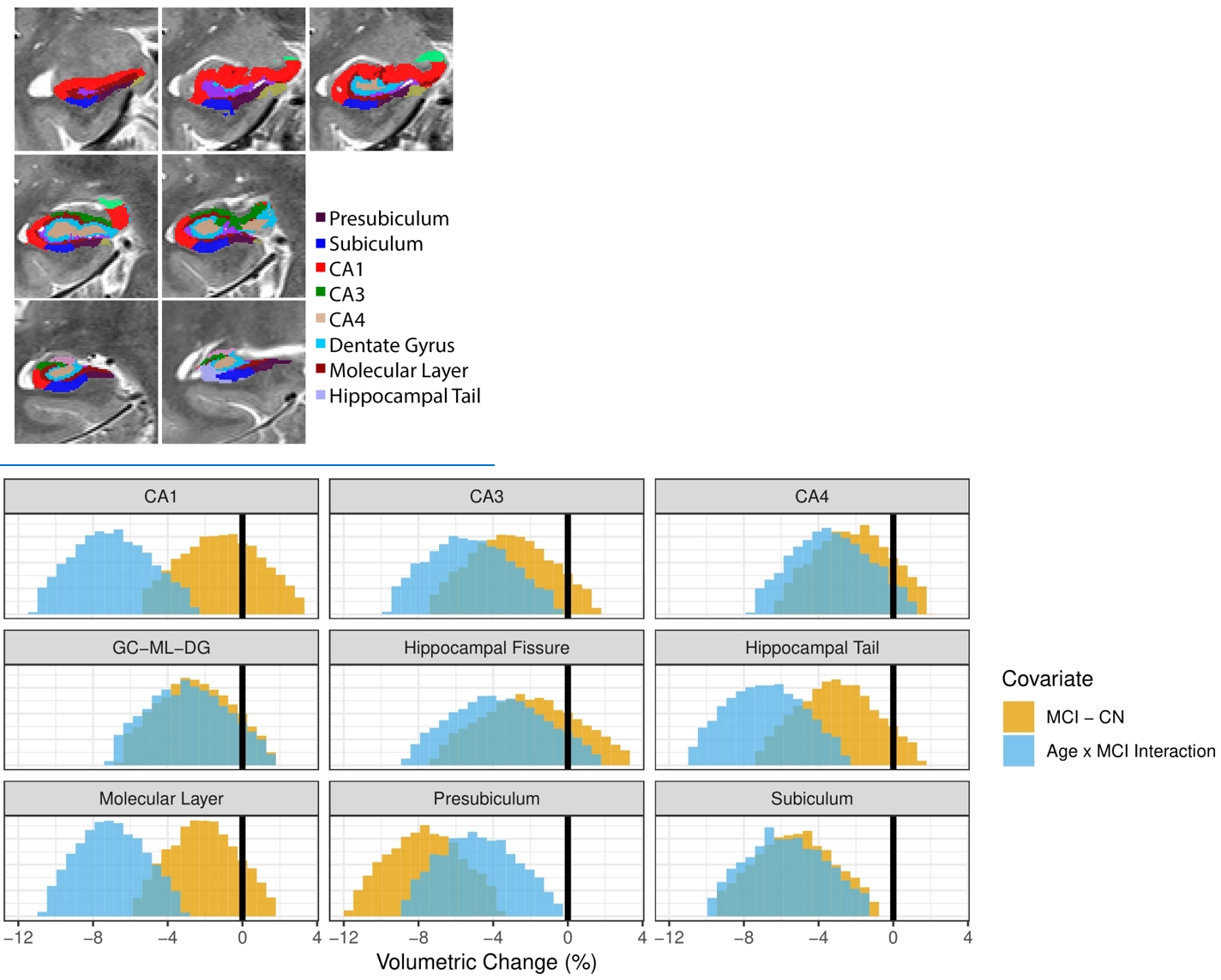
Top: Sample
hippocampal subfield segmentation results. Bottom: Distribution of Bayesian
regression of hippocampal subfield volumes examining MCI effects and age x MCI
interaction effects. Distribution tails crossing zero are non-significant. Presubiculum and subiculum volumes were
significantly decreased in MCI. CA1, CA3, hippocampal tail, molecular layer,
presubiculum, and subiculum volumes were negatively associated with age x MCI
interaction.