Gaurav Verma1, Rebecca Emily Feldman2, and Priti Balchandani1
1Radiology, Icahn School of Medicine at Mount Sinai, New York, NY, United States, 2Medical Physics, University of British Columbia, Kelowna, BC, Canada
1Radiology, Icahn School of Medicine at Mount Sinai, New York, NY, United States, 2Medical Physics, University of British Columbia, Kelowna, BC, Canada
An
echo-planar spectroscopic imaging (EPSI) sequence has been demonstrated combining semi-adiabatic spatial-spectral pulses and echo-planar readout to facilitate fast magnetic resonance spectroscopic imaging (MRSI) at ultra-high field (7T)
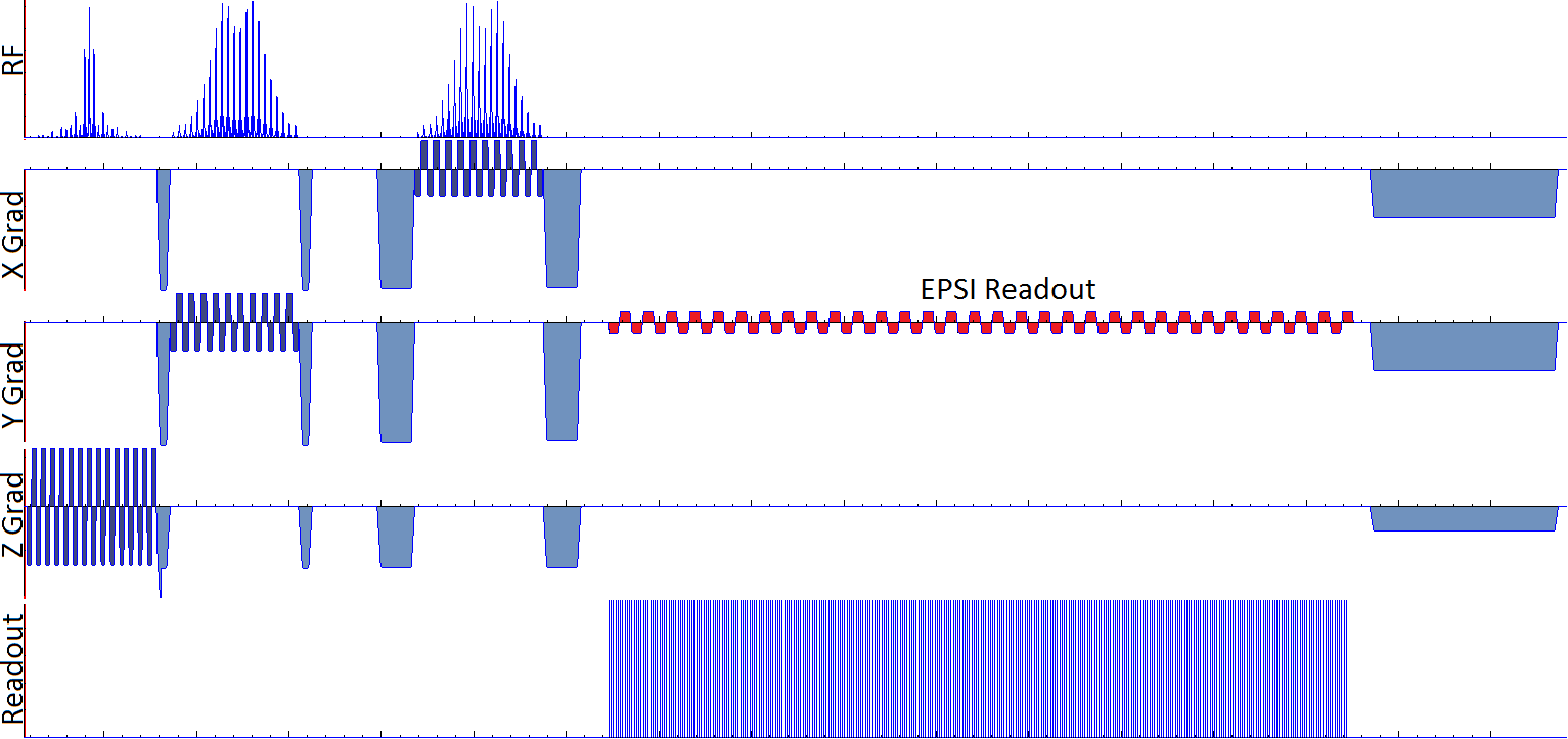
Figure 1: Pulse sequence diagram of the EPSI sequence showing semi-adiabatic point-resolved spectroscopy (PRESS) sequence and bi-polar echo-planar readout.
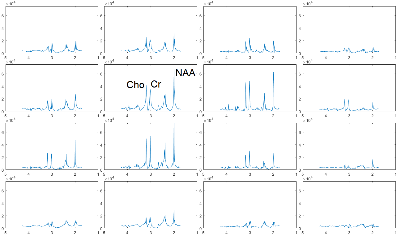
Figure 4: Fully-reconstructed data showing spatially-resolved spectra with peaks of Creatine (Cr), Choline (Cho) and N-acetyl aspartate (NAA) visible and labeled in one spectrum. Horizontal axes are in parts-per-million (ppm) and vertical axes in arbitrary units representing signal amplitude.