Xiaoxuan He1, Edward J. Auerbach1, Michael Garwood1, Naoharu Kobyashi1, Alireza Sadeghi‐Tarakameh1, Yigitcan Eryaman1, Xiaoping Wu1, and Gregory J. Metzger1
1Center for Magnetic Resonance Research, University of Minnesota, Minneapolis, MN, United States
1Center for Magnetic Resonance Research, University of Minnesota, Minneapolis, MN, United States
A parallel
transmit optimized 3D spatial-spectral pulse is developed for spectroscopic
imaging for brain studies at 10.5T with reduced SAR, intrinsic water suppression
and field inhomogeneity mitigation. Phantom studies are used to compare the new
method with a conventional approach.
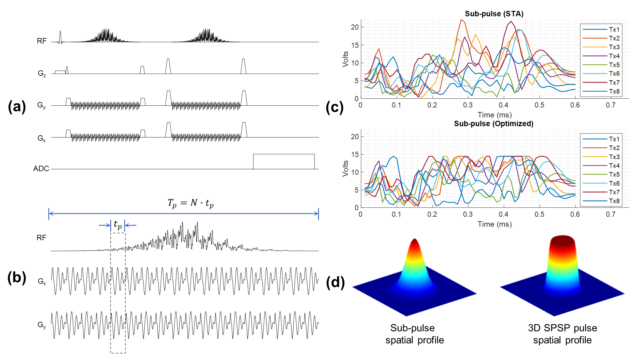
Figure 1. The
proposed spectroscopy acquisition at 10.5T using (a) a double spin-echo
sequence with (b) 3D SPSP adiabatic pulses for refocusing. The design of the 3D
SPSP pulse was shown in (b), where a train of pTx spiral sub-pulses are
modulated by an adiabatic envelope. With pTx, the peak amplitude can be
effectively reduced and hard constrained by optimal control method as shown in
(c). To further increase the spectral bandwidth at 10.5T, an inhomogeneous
spatial profile can be used for the sub-pulse while still achieving a
homogeneous inversion by the 3D SPSP pulses as shown in (d).
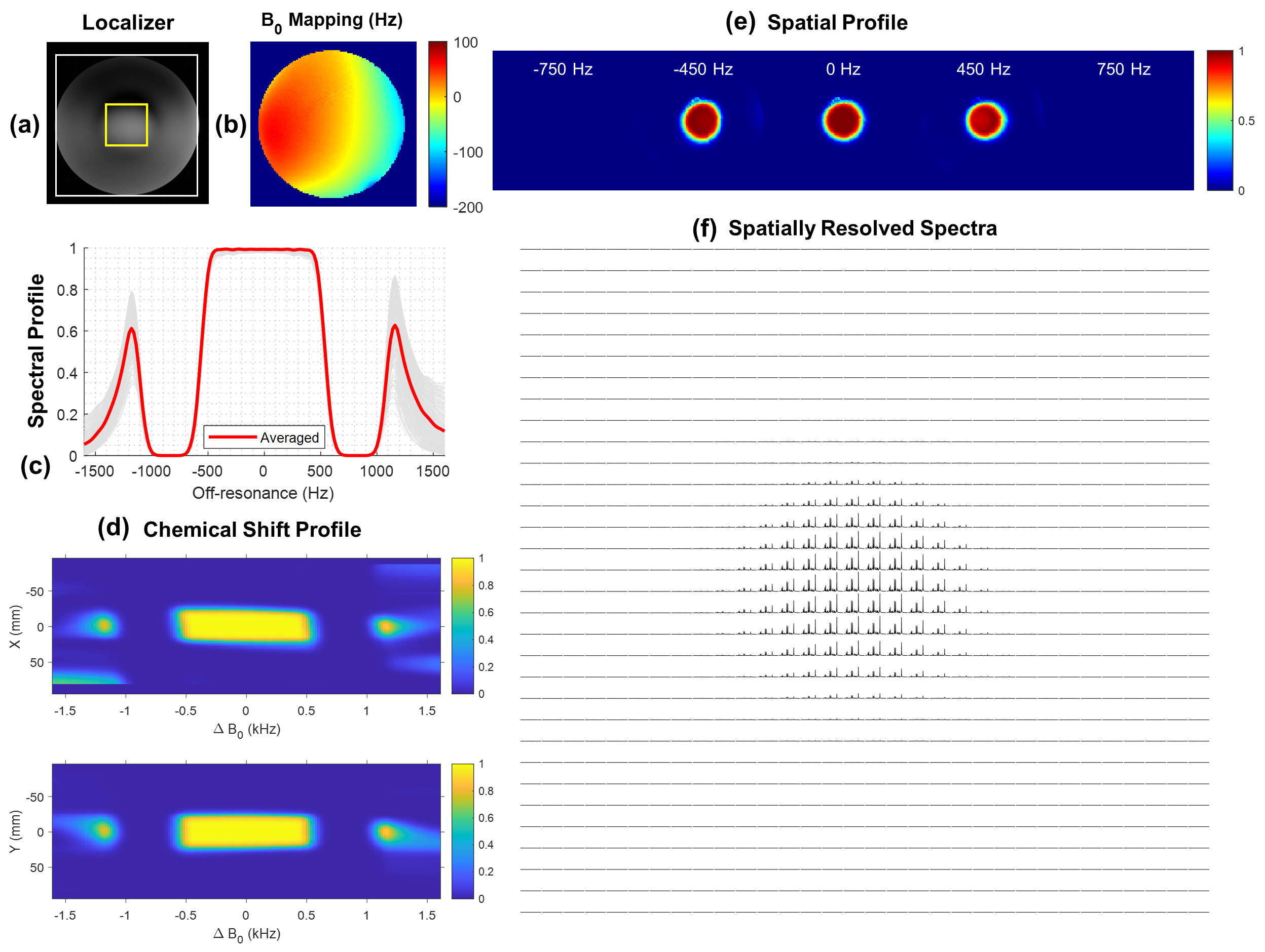
Figure 2. A
brief summary of the 3D SPSP pulse performance. The phantom setup was shown in (a),
where FOV and VOI were indicated by the white and yellow box. The B0 mapping
was shown in (b). By simulation, the pulse provided an approximately 920 Hz for
the 95% passband along with stopband at ±750 Hz for water and lipid suppression
as shown in (c), with overall mild chemical shift displacement errors as shown
in (d), (e). The spatially resolved spectra acquired with the proposed method
as shown in (f) matched with the simulated profiles.