Olivia E Rowe1, Rangaprakash Deshpande1, Akila Weerasekera1, Christopher Stephen2,3, Robert L Barry1,4, Florian Eichler3,5, and Eva-Maria Ratai1
1Athinoula A. Martinos Center for Biomedical Imaging, Department of Radiology, Massachusetts General Hospital and Harvard Medical School, Charlestown, MA, United States, 2Movement Disorders Division and Ataxia Center, Department of Neurology, Massachusetts General Hospital and Harvard Medical School, Boston, MA, United States, 3Center for Rare Neurological Diseases, Department of Neurology, Massachusetts General Hospital and Harvard Medical School, Boston, MA, United States, 4Harvard-Massachusetts Institute of Technology Health Sciences & Technology, Cambridge, MA, United States, 5Leukodystrophy Clinic, Department of Neurology, Massachusetts General Hospital and Harvard Medical School, Boston, MA, United States
1Athinoula A. Martinos Center for Biomedical Imaging, Department of Radiology, Massachusetts General Hospital and Harvard Medical School, Charlestown, MA, United States, 2Movement Disorders Division and Ataxia Center, Department of Neurology, Massachusetts General Hospital and Harvard Medical School, Boston, MA, United States, 3Center for Rare Neurological Diseases, Department of Neurology, Massachusetts General Hospital and Harvard Medical School, Boston, MA, United States, 4Harvard-Massachusetts Institute of Technology Health Sciences & Technology, Cambridge, MA, United States, 5Leukodystrophy Clinic, Department of Neurology, Massachusetts General Hospital and Harvard Medical School, Boston, MA, United States
Late-onset GM2-Gangliosidoses (LOGG), which are rare lysosomal storage disorders and include Tay-Sachs disease (LOTS) and Sandhoff disease (LOSD), have notable structural and metabolic effects on the cerebellum.
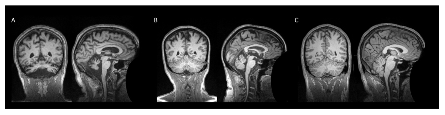
Figure 1: T1-weighted MR images showing cerebellar atrophy. Defaced coronal and sagittal MRI slices (MNI space: x=0) of (A) patient presenting with late-onset Tay-Sachs disease showing profound cerebellar atrophy, (B) patient presenting with late-onset Sandhoff disease, and (C) healthy control.
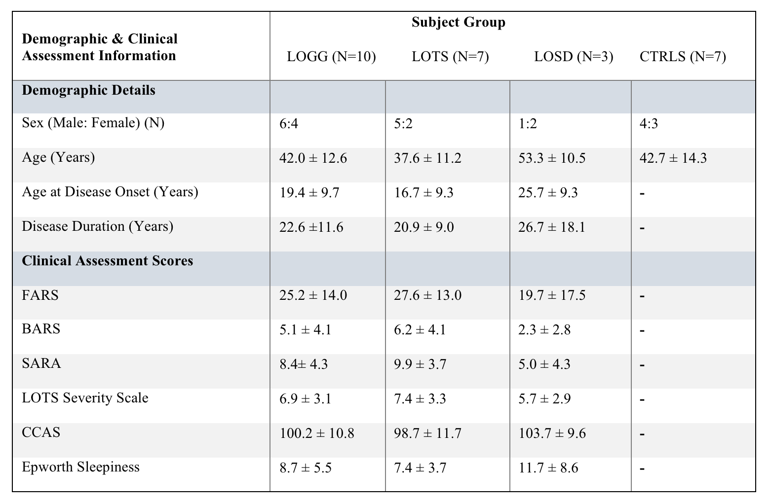
Table 1: Participant demographics and clinical assessment scores reported as mean ± standard deviation.