Dunja Simicic1,2,3, Jessie Julie Mosso1,2,3, Thanh Phong Lê3,4, Ruud B. van Heeswijk5, Ileana Ozana Jelescu1,2, and Cristina Cudalbu1,2
1CIBM Center for Biomedical Imaging, Lausanne, Switzerland, 2Animal Imaging and Technology, EPFL, Lausanne, Switzerland, 3Laboratory of Functional and Metabolic Imaging, EPFL, Lausanne, Switzerland, 4Geneva School of Health Sciences, HES-SO University of Applied Sciences and Arts Western Switzerland, Geneva, Switzerland, 5Department of Radiology, Lausanne University Hospital (CHUV) and University of Lausanne (UNIL), Lausanne, Switzerland
1CIBM Center for Biomedical Imaging, Lausanne, Switzerland, 2Animal Imaging and Technology, EPFL, Lausanne, Switzerland, 3Laboratory of Functional and Metabolic Imaging, EPFL, Lausanne, Switzerland, 4Geneva School of Health Sciences, HES-SO University of Applied Sciences and Arts Western Switzerland, Geneva, Switzerland, 5Department of Radiology, Lausanne University Hospital (CHUV) and University of Lausanne (UNIL), Lausanne, Switzerland
MRSI is a powerful tool for the non-invasive simultaneous mapping of metabolic profiles at multiple spatial positions. Aim of the present study was to implement an improved denoising technique (MP-PCA) on high resolution MRSI data acquired at 9.4T in the rat-brain.
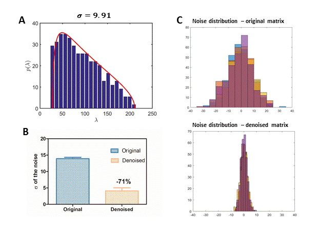
Figure 1. a) Fit of
Marchenko-Patstur distribution to the lowest eigenvalues from PCA. B) Mean
value of the standard deviation of noise (last 300 points of the FID) from all
120 spectra before and after denoising (upper and lower graphs, respectively). c)
The noise distribution of 6 random selected spectra before and after denoising.
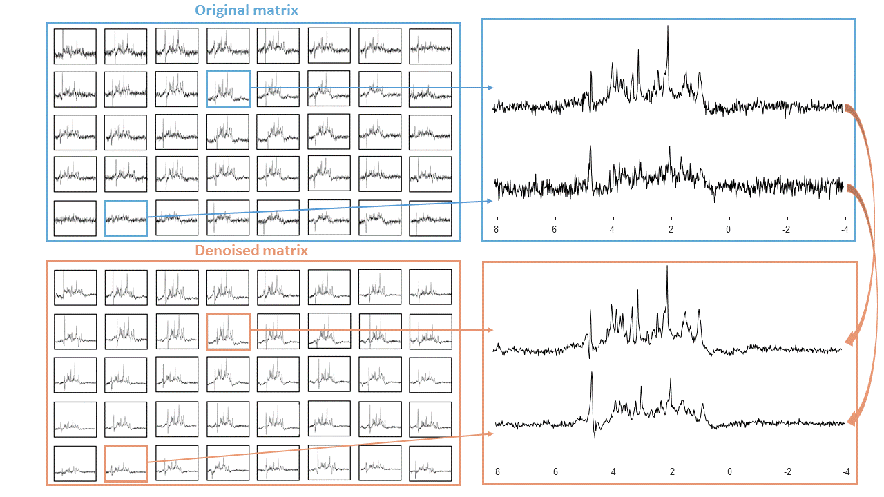
Figure 2. A sub-set
(5x8) of the full image matrix demonstrates the quality of the spectra before
and after denoising.