Benjamin Ades-Aron1, Hong-Hsi Lee1, Heidi Schambra2, Dmitry S. Novikov1, Els Fieremans1, and Timothy Shepherd1
1Radiology, NYU School of Medicine, New York, NY, United States, 2Neurology, NYU Langone, New York, NY, United States
1Radiology, NYU School of Medicine, New York, NY, United States, 2Neurology, NYU Langone, New York, NY, United States
We describe and
evaluate a novel combination of Fast Gray Matter Acquisition T1 Inversion
Recovery (FGATIR) denoising with deep learning and super-resolution techniques to improve the
accuracy of small internal brainstem structure segmentation on advanced
diffusion MRI data
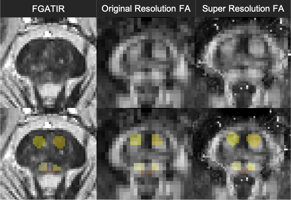
Co-registered
axial images of mid-pons using FGATIR, standard and super-resolution fractional
anisotropy with overlaid segmentations of corticospinal tract (PON; dark
yellow), pontine reticular formation (PRF; light yellow) and medial
longitudinal fasciculus (MLF; red) created by neuroanatomy expert using FGATIR
contrast propagated to the diffusion data. The round shape of the PON
segmentation is better preserved with super-resolution. There is less CSF
contamination of the MLF from the 4th ventricle with super-resolution.
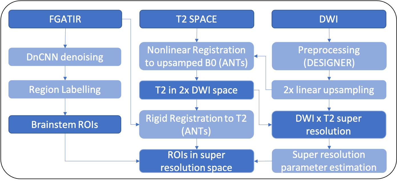
Overview of the pipeline. Images are labelled as
solid blue blocks and processing steps are labelled as light blue blocks. The
pipeline relies on denoised FGATIR data that undergoes diffeomorphic
registration through a 3D T2 weighted image to a super-resolution enhanced
diffusion weighted dataset.