Maria Guadalupe Garcia Gomar1, Kavita Singh1, and Marta Bianciardi1
1Radiology, Athinoula A. Martinos Center for Biomedical Imaging, Massachusetts General Hospital and Harvard Medical School, Boston, MA, United States, Charlestown, MA, United States
1Radiology, Athinoula A. Martinos Center for Biomedical Imaging, Massachusetts General Hospital and Harvard Medical School, Boston, MA, United States, Charlestown, MA, United States
Pontine and medullary brainstem nuclei play a crucial role in arousal and sleep, yet their study in humans is challenging due to limited resolution and contrast of conventional MRI. We generated an in-vivo probabilistic structural atlas and connectome of brainstem nuclei using 7 Tesla MRI.
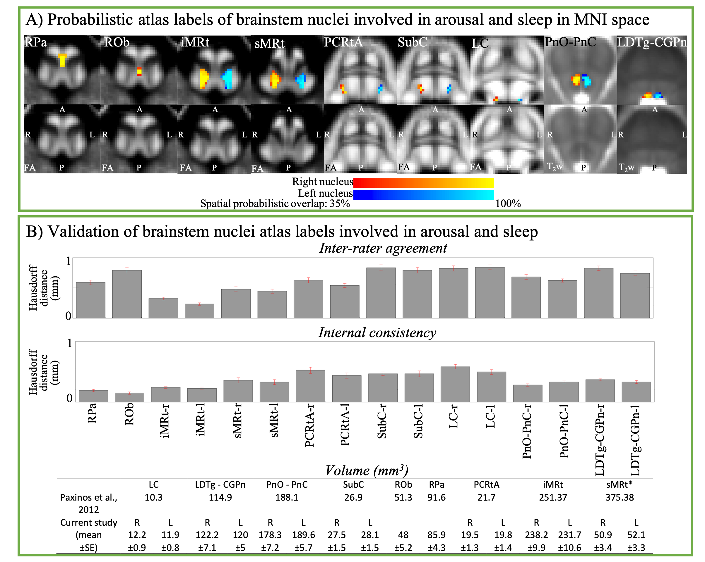
Figure1: A) Probabilistic (n = 12) atlas label of nine brainstem nuclei overlaid on the group averaged FA or T2w image. We observed very good (up to 100 %) spatial agreement of labels across subjects indicating the feasibility of delineating these nuclei based on 7 Tesla MRI contrast and neighborhood rules7. B) For each nucleus, the inter-rater agreement and internal consistency of nuclei labels were below (p < 0.05) the linear imaging resolution (1.1 mm), thus validating the nuclei atlas. Except for sMRt, volumes of nuclei labels in-vivo did not differ (p < 0.05) from literature values7.
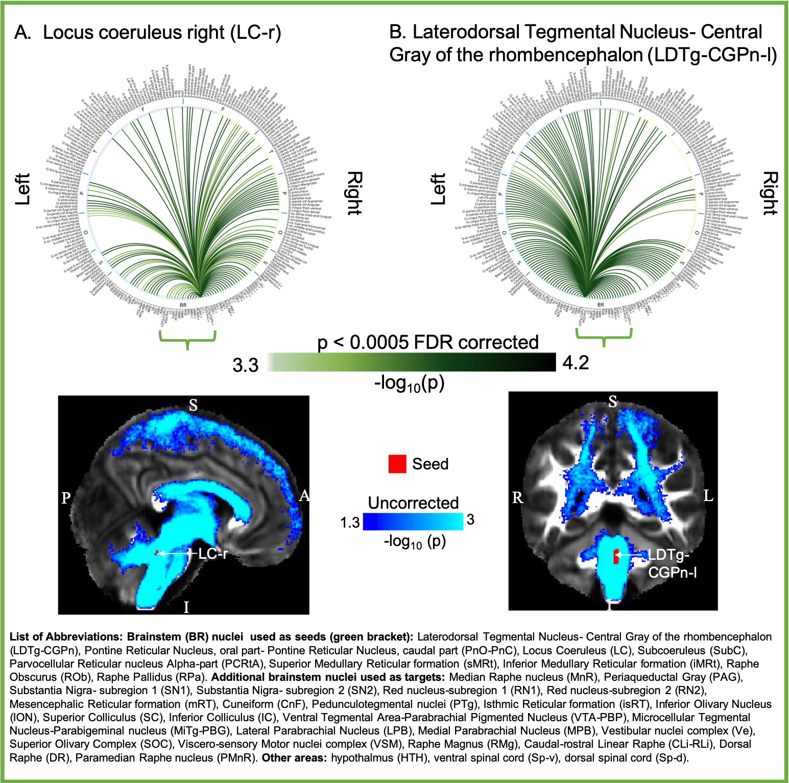
Figure 4. Top) Structural connectome and Bottom) streamline density (n=19) of two pontine brainstem nuclei: A. right LC-r and B. left LDTg-CGPn (-log10(p-value) displayed, Wilcoxon test). LC showed connectivity with bilateral iMRt, sMRt, HTH, striatum and widespread cortical connectivity including limbic areas in line with previous studies2. LDTg-CGPn, described previously as the most extensive group of the rostral braintem2 showed connectivity with HTH, VTA, PAG, basal forebrain, frontal and limbic areas as expected from previous literature2,7.
