Iain P Bruce1, Yixin Ma1, and Allen W Song1
1Brain Imaging and Analysis Center, Duke University, Durham, NC, United States
1Brain Imaging and Analysis Center, Duke University, Durham, NC, United States
To delineate abnormalities in cortical microstructures, this study presents a technique to accurately and effectively observe the cortical depth dependence of diffusion anisotropy and diffusivity in-vivo using sub-millimeter isotropic resolution diffusion tensor imaging.
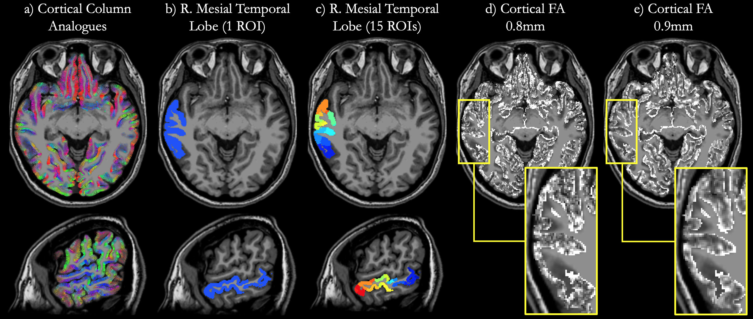
Figure 2. Cortical column analogues (a) derived from surface normal vectors spanning between white matter and the pial surface. For analysis, the right mesial temporal lobe is shown as a single region (b) and partitioned into 15 regions (c). To observe depth dependence of diffusion anisotropy, cortical FA maps from DTI data acquired at 0.8mm (d) and 0.9mm (e) are projected onto the column analogues in each region of interest.
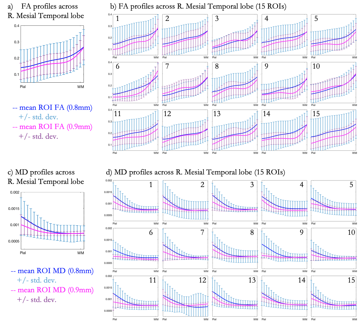
Figure 3. Cortical profiles of fractional anisotropy (FA) projected onto all column analogues in the right mesial temporal lobe (a) and in each of 15 sub-regions (b). Profiles from 0.8mm (blue) and 0.9mm (pink) DTI data show an increase in FA with cortical depth from the pial surface (PS) to the white matter (WM) boundary, with local extrema in the middle. Similar profiles for mean diffusivity (MD) (c-d) show a reduction in diffusivity across the cortex from the PS to the WM boundary, with no local extrema.