Sebastian Papazoglou1, Mohammad Ashtarayeh1, Martina F. Callaghan2, Mark D. Does3,4,5,6, and Siawoosh Mohammadi1,7
1Department of Systems Neurosciences, University Medical Center Hamburg-Eppendorf, Hamburg, Germany, 2Wellcome Centre for Human Neuroimaging, UCL Queen Square Institute of Neurology, University College London, London, United Kingdom, 3Department of Biomedical Engineering, Vanderbilt University, Nashville, TN, United States, 4Institute of Imaging Science, Vanderbilt University Medical Center, Nashville, TN, United States, 5Department of Radiology and Radiological Sciences, Vanderbilt University Medical Center, Nashville, TN, United States, 6Department of Electrical Engineering, Vanderbilt University, Nashville, TN, United States, 7Department of Neurophysics, Max Planck Institute for Human Cognitive and Brain Sciences, Leipzig, Germany
1Department of Systems Neurosciences, University Medical Center Hamburg-Eppendorf, Hamburg, Germany, 2Wellcome Centre for Human Neuroimaging, UCL Queen Square Institute of Neurology, University College London, London, United Kingdom, 3Department of Biomedical Engineering, Vanderbilt University, Nashville, TN, United States, 4Institute of Imaging Science, Vanderbilt University Medical Center, Nashville, TN, United States, 5Department of Radiology and Radiological Sciences, Vanderbilt University Medical Center, Nashville, TN, United States, 6Department of Electrical Engineering, Vanderbilt University, Nashville, TN, United States, 7Department of Neurophysics, Max Planck Institute for Human Cognitive and Brain Sciences, Leipzig, Germany
All DWI-models improved with additional calibration. Models with fixed
diffusivities benefited efficiently from an additional scaling, while
WMTI model benefited by an additional offset. Without calibration the axonal compartment
biomarker from NODDIDTI could best explain the data.
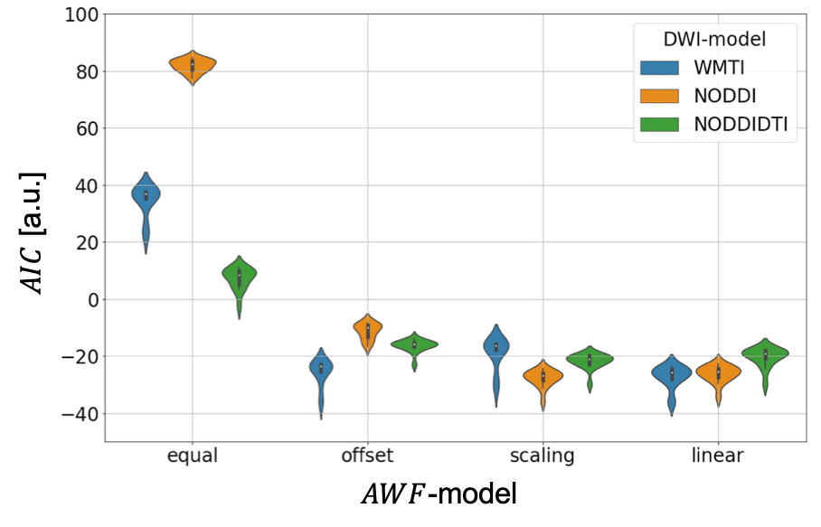
Figure 3: for the
validated DWI-model + $$$AWF$$$-model. NODDIDTI has the largest $$$AIC$$$ out-of-the-box, i.e. with no adjustment
between biomarker and axonal water fraction. While in general, all DWI models
benefit significantly from additional calibration parameters, WMTI appears to
benefit the most from an additional offset and both NODDI-models benefit from
an additional scaling. The introduction of a fully linear calibration does not
lead to significant further improvements in terms of $$$AIC$$$.
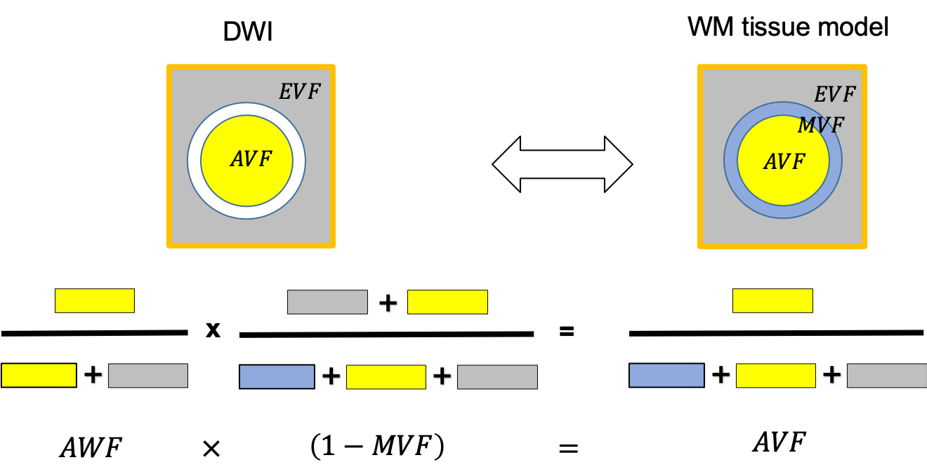
Figure 1: Schematic of the relationship between three-compartment WM tissue model and the DWI. Here, WM is assumed to be composed of three tissue compartments: axonal ($$$AVF$$$), myelin ($$$MVF$$$), and extracellular volume fraction ($$$EVF$$$). Since myelin is DWI-invisible, the axonal compartment accessible via DWI is the axonal water fraction ($$$AWF$$$). Hence DWI-based biomarkers for the axonal compartment have to be rescaled by to yield an estimate
$$f_A=(1-\mu)AWF\qquad (1)$$
of the $$$AVF$$$. This information has to be acquired from another technique.