Luke Joel Edwards1, Evgeniya Kirilina1,2, Carsten Jäger1,3, Kirsten Garus4, Markus Cremer4, Katrin Amunts4,5, and Nikolaus Weiskopf1,6
1Department of Neurophysics, Max Planck Institute for Human Cognitive and Brain Sciences, Leipzig, Germany, 2Center for Cognitive Neuroscience Berlin, Freie Universität Berlin, Berlin, Germany, 3Paul Flechsig Institute of Brain Research, Leipzig University, Leipzig, Germany, 4Institute of Neuroscience and Medicine, Research Centre Jülich, Jülich, Germany, 5C. and O. Vogt Institute for Brain Research, Heinrich Heine University Düsseldorf, Düsseldorf, Germany, 6Felix Bloch Institute for Solid State Physics, Leipzig University, Leipzig, Germany
1Department of Neurophysics, Max Planck Institute for Human Cognitive and Brain Sciences, Leipzig, Germany, 2Center for Cognitive Neuroscience Berlin, Freie Universität Berlin, Berlin, Germany, 3Paul Flechsig Institute of Brain Research, Leipzig University, Leipzig, Germany, 4Institute of Neuroscience and Medicine, Research Centre Jülich, Jülich, Germany, 5C. and O. Vogt Institute for Brain Research, Heinrich Heine University Düsseldorf, Düsseldorf, Germany, 6Felix Bloch Institute for Solid State Physics, Leipzig University, Leipzig, Germany
Ultra-strong diffusion gradients, novel gradient nonlinearity correction, and excellent tissue quality of the post mortem brain enabled collection of data which will be used for validation of higher order diffusion models and tractography.
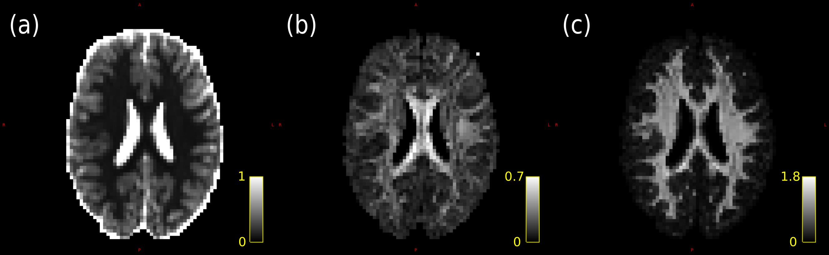
Figure 1: Diffusion kurtosis maps. (a) Mean diffusivity (µm2/ms). (b) Fractional anisotropy. (c) Mean kurtosis.
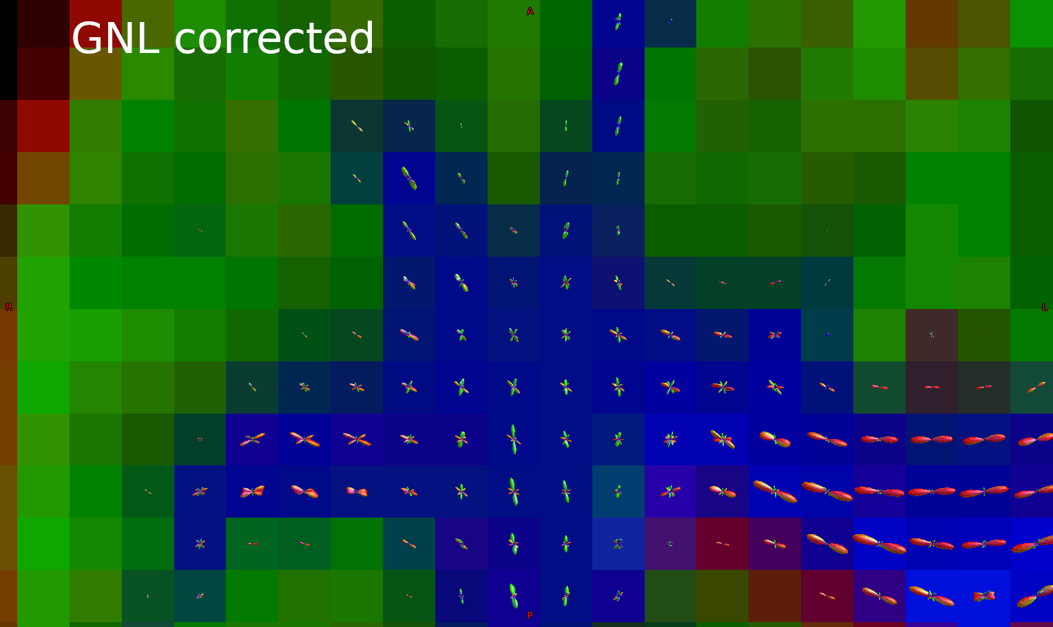
Figure 3: Animated GIF showing fODFs with and without GNL correction in the frontal lobe. Subtle but distinct differences can be seen after correction.