Mohammad Ashtarayeh1, Laurin Mordhorst1, Maria Morozova2,3, Tobias Streubel1,2, Jan Malte Oeschger1, Joao Periquito4, Andreas Pohlmann4, Henriette Rusch3, Carsten Jäger2, Thoralf Niendorf4, Nikolaus Weiskopf2,5, Markus Morawski2,3, and Siawoosh Mohammadi1,2
1Department of Systems Neurosciences, University Medical Center Hamburg-Eppendorf, Hamburg, Germany, 2Department of Neurophysics, Max Planck Institute for Human Cognitive and Brain Sciences, Leipzig, Germany, 3Paul Flechsig Institute of Brain Research, University of Leipzig, Leipzig, Germany, 4Berlin Ultrahigh Field Facility (B.U.F.F.), Max-Delbrueck-Center for Molecular Medicine in the Helmholtz Association, Berlin, Germany, 5Felix Bloch Institute for Solid State Physics, Faculty of Physics and Earth Sciences, Leipzig University, Leipzig, Germany
1Department of Systems Neurosciences, University Medical Center Hamburg-Eppendorf, Hamburg, Germany, 2Department of Neurophysics, Max Planck Institute for Human Cognitive and Brain Sciences, Leipzig, Germany, 3Paul Flechsig Institute of Brain Research, University of Leipzig, Leipzig, Germany, 4Berlin Ultrahigh Field Facility (B.U.F.F.), Max-Delbrueck-Center for Molecular Medicine in the Helmholtz Association, Berlin, Germany, 5Felix Bloch Institute for Solid State Physics, Faculty of Physics and Earth Sciences, Leipzig University, Leipzig, Germany
Our results confirms that large-scale light microscopy is an improved reference standard compared to the manually-labeled electron microscopy for validating MRI-based axon radius index.
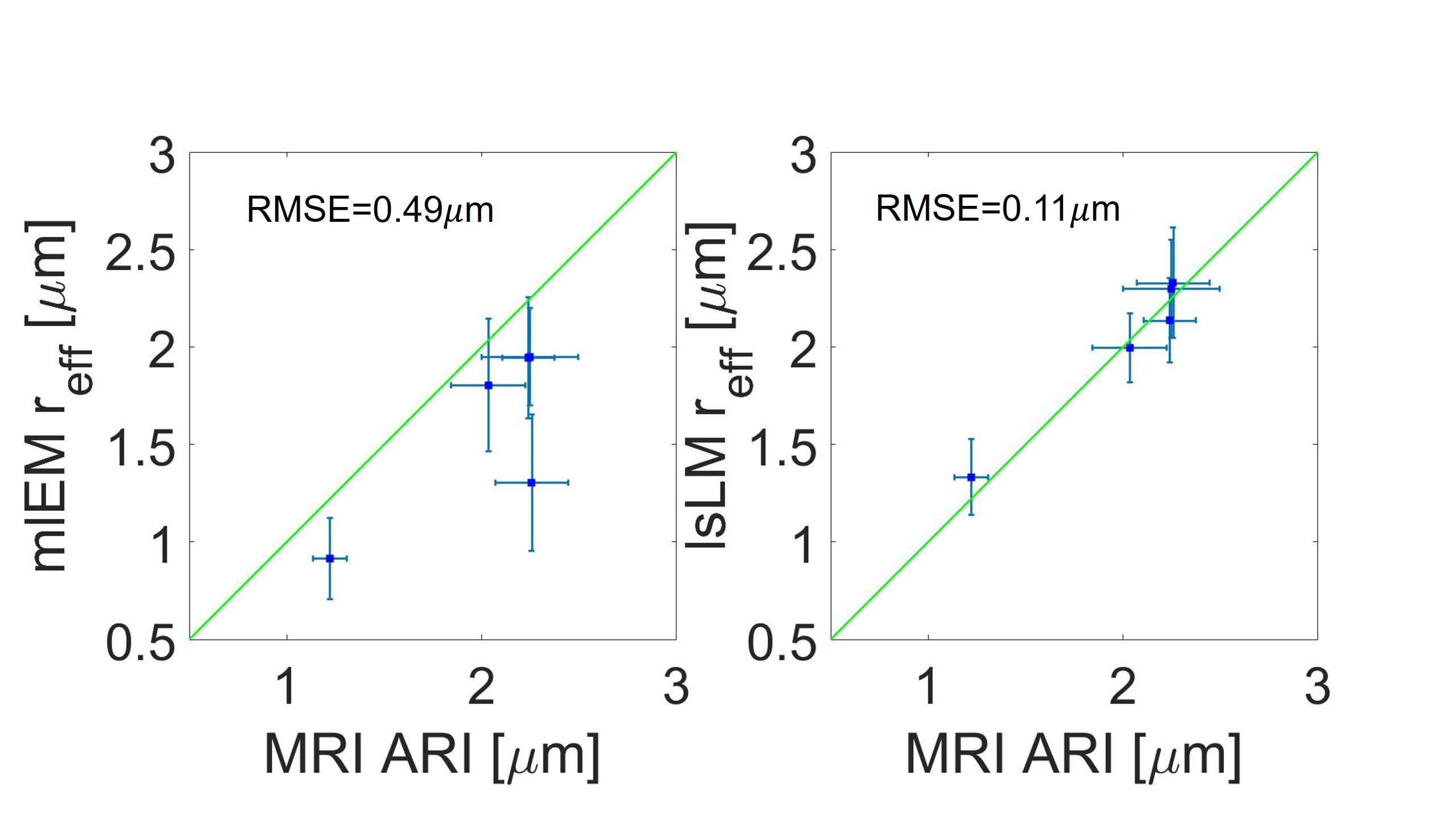
Fig. 4: Comparing MRI-based axon radius index (ARI) estimation
with the histological manually labeled electron microscopy (mlEM) and
automatically labelled large-scale light microscopy (lsLM) for five region of
interests. (a): Depicted is a scatter plot of the estimated ARIs from MRI
against the effective axon radius from mlEM. (b): Depicted is scatter plot of
the estimated ARIs from MRI against the effective axon radius from lsLM. The RMSE in mlEM is 0.49 µm whereas it is 0.1 µm in lsLM.
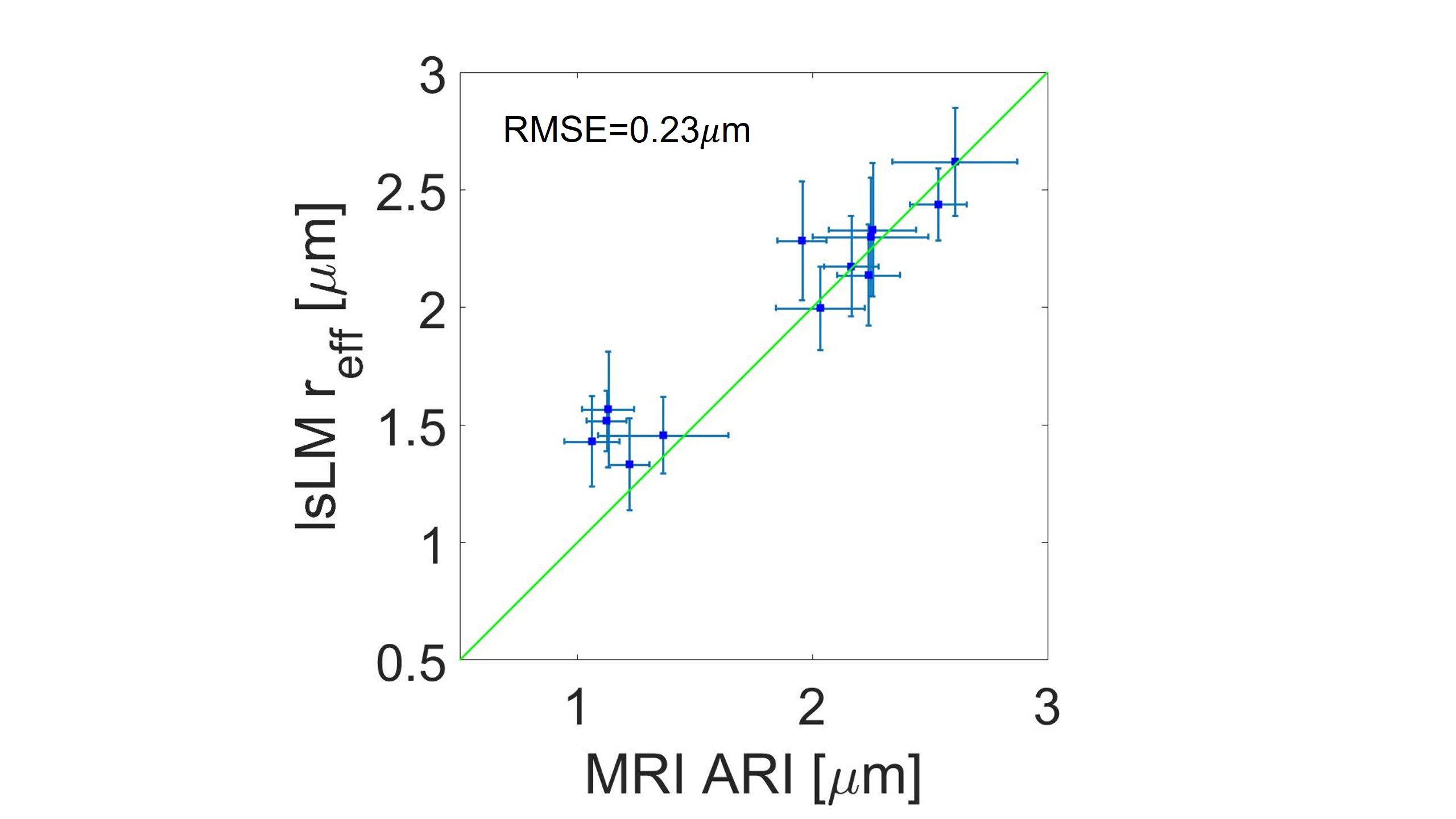
Fig. 5:
Assessing the accuracy of MRI-based axon radius index (ARI) estimation by
comparison with large-scale light microscopy (lsLM) across eighteen regions of
interests in the corpus callosum. From the eighteen investigated regions, only thirteen
were used in this analysis. The remaining five regions were insufficient tissue
or data quality, either in lsLM or MRI.