Sidsel Winther1,2, Mariam Andersson1,2, Henrik Lundell2, and Tim Dyrby1,2
1DTU Compute, Technical University of Denmark, Kongens Lyngby, Denmark, 2Danish Research Center for Magnetic Resonance, Functional and Diagnostic Imaging and Research, Copenhagen University Hospital Hvidovre, Hvidovre, Denmark
1DTU Compute, Technical University of Denmark, Kongens Lyngby, Denmark, 2Danish Research Center for Magnetic Resonance, Functional and Diagnostic Imaging and Research, Copenhagen University Hospital Hvidovre, Hvidovre, Denmark
Susceptibility of myelin induces morphology- and orientation-dependent perturbations at the microstructural scale of the B_0-field. We show that a consequence of this is a fiber-orientation-dependent bias of the DWI-signal across white matter regions.
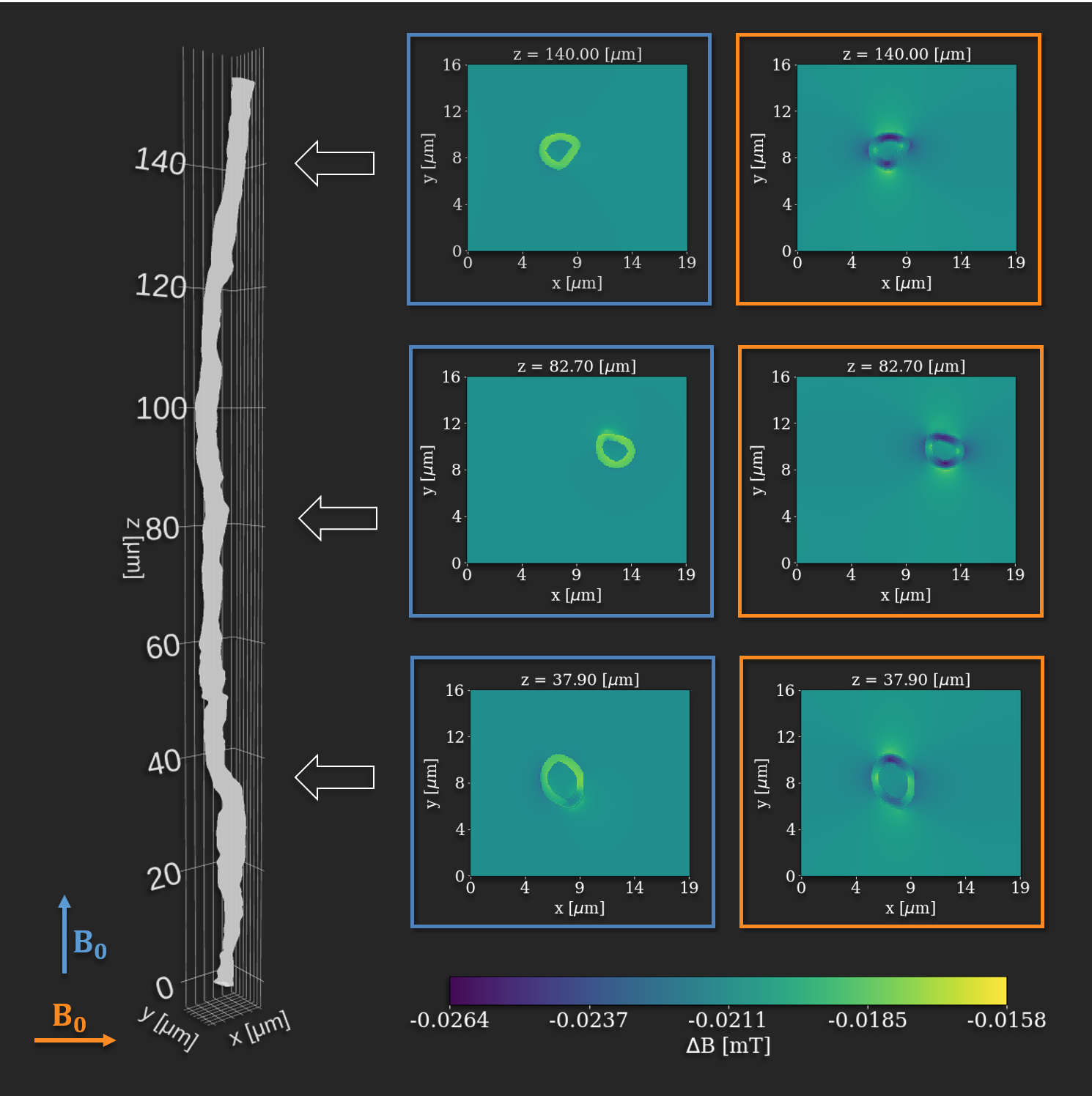
Figure 1: Left: Axon from the corpus callosum of a vervet monkey brain (segmented from 3D x-ray nano-holotomography). Right: Cross-sections of the perturbations $$$\Delta B$$$ of an applied field of 7 T computed with 0.1 μm isotropic resolution with an in-house software for parallel (blue) and perpendicular (orange) orientation w.r.t. the field. The field is most affected when fibers are oriented perpendicular to the field. The gradients are stronger perpendicular to the axon than parallel to it.
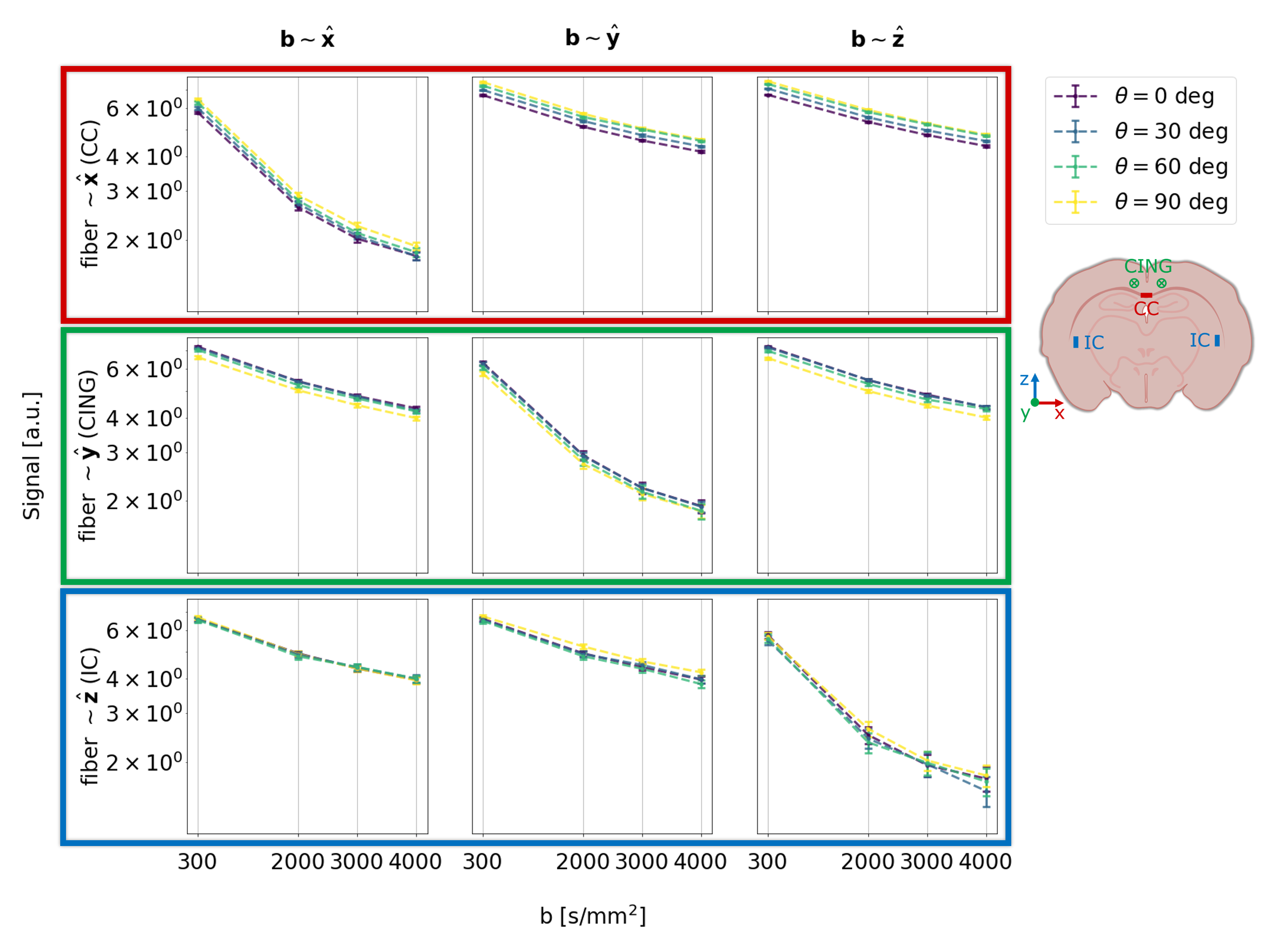
Figure 4: 1st, 2nd, and 3rd row shows the signal from CC, CING, and IC, respectively. 1st, 2nd, and 3rd column show the signal from the b-vectors closest to x, y, and z, respectively. Hence, the diagonal shows the signal measured parallel to the fibers, and the off-diagonal shows signal measured perpendicular to the fibers. Each point represents the average of all voxels of interest. Number of voxels varied across scans: 681 to 816, 209 to 283, and 64 to 93 for CC, CING, and IC, respectively.