Noam Omer1, Ella Wilczynski1, Neta Stern1, Tamar Blumenfeld-Katzir1, Meirav Galun2, and Noam Ben-Eliezer1,3,4,5,6
1Department of Biomedical Engineering, Tel-Aviv University, Tel-Aviv, Israel, 2Department of Computer Science and Applied Mathematics, Weitzman institute of science, Rehovot, Israel, 3Department of Orthopedics, Shamir Medical Center, Zerifin, Israel, 4Sackler Faculty of Medicine, Tel-Aviv University, Tel-Aviv, Israel, 5Sagol School of Neuroscience, Tel-Aviv University, Tel-Aviv, Israel, 6Center for Advanced Imaging Innovation and Research (CAI2R), New-York University Langone Medical Center, New York, NY, United States
1Department of Biomedical Engineering, Tel-Aviv University, Tel-Aviv, Israel, 2Department of Computer Science and Applied Mathematics, Weitzman institute of science, Rehovot, Israel, 3Department of Orthopedics, Shamir Medical Center, Zerifin, Israel, 4Sackler Faculty of Medicine, Tel-Aviv University, Tel-Aviv, Israel, 5Sagol School of Neuroscience, Tel-Aviv University, Tel-Aviv, Israel, 6Center for Advanced Imaging Innovation and Research (CAI2R), New-York University Langone Medical Center, New York, NY, United States
A novel data-driven approach for multi T2-component analysis produces reliable estimation of myelin content in
vivo. Validation shows excellent agreement between this technique and ground
truth immunohistochemical staining of myelin basic protein.
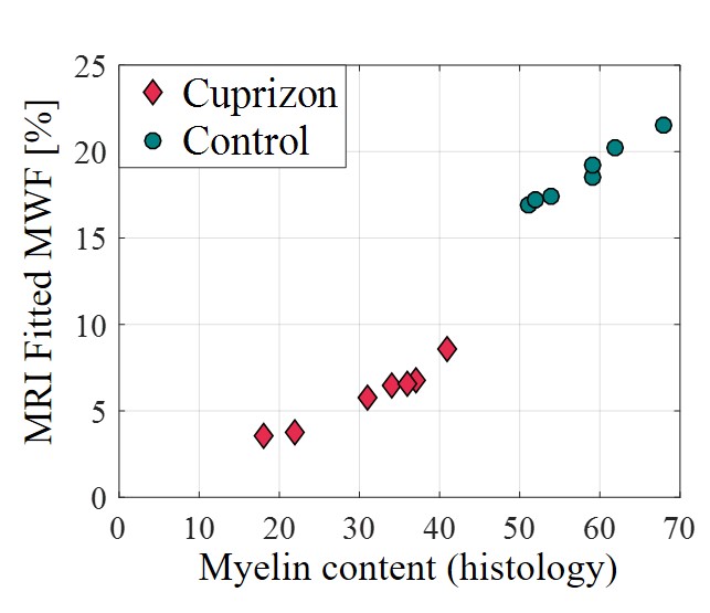
Figure 4. Scatter
plot
of estimated myelin content at the medial croups callosum of demyelinated mice
vs. healthy controls. Values were calculated based on immunohistochemical
(IHC) staining vs. MRI based myelin water fractions (MWFs) fitted using the
suggested mcT2 technique. Good agreement is shown between the two techniques,
attesting to the ability to classify the two groups and detect demyelination
based on MRI derived MWF values.
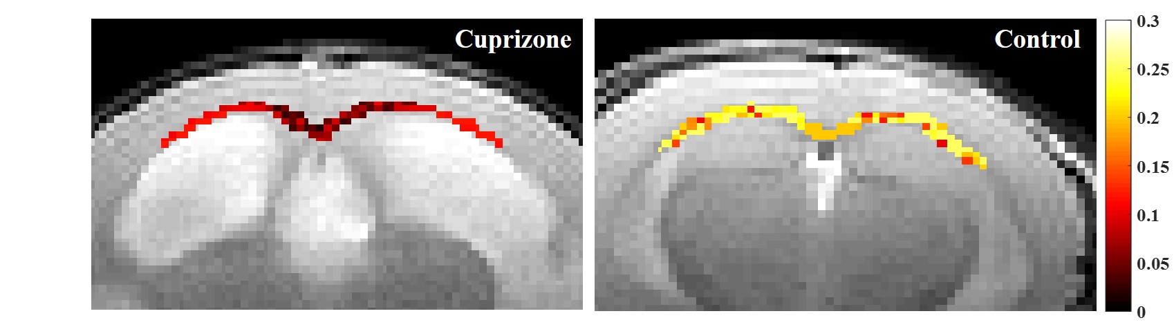
Figure 3. Example myelin water fraction (MWF) maps based on MRI derived MWF values. MWF values are presented along the croups callosum (CC) for cuprizone (left) and (right) control mice showing lower values for the cuprizone mouse. Maps are shown on top the T2-wighted image (5th echo) obtained using multi-echo-spic-echo MRI protocol and with the same colormap.