Xinnan Li1, Daisuke Sawamura2, Hiroyuki Hamaguchi1, Yuta Urushibata3, Thorsten Feiweier4, Keita Ogawa5, and Khin Khin Tha1,6
1Department of Biomarker Imaging Science, Graduate School of Biomedical Science and Engineering, Hokkaido University, Sapporo, Japan, 2Department of Functioning and Disability, Faculty of Health Sciences, Hokkaido University, Sapporo, Japan, 3Siemens Healthcare K.K., Tokyo, Japan, 4Siemens Healthcare GmbH, Erlangen, Germany, 5Department of Rehabilitation, Hokkaido University Hospital, Sapporo, Japan, 6Global Center for Biomedical Science and Engineering, Faculty of Medicine, Hokkaido University, Sapporo, Japan
1Department of Biomarker Imaging Science, Graduate School of Biomedical Science and Engineering, Hokkaido University, Sapporo, Japan, 2Department of Functioning and Disability, Faculty of Health Sciences, Hokkaido University, Sapporo, Japan, 3Siemens Healthcare K.K., Tokyo, Japan, 4Siemens Healthcare GmbH, Erlangen, Germany, 5Department of Rehabilitation, Hokkaido University Hospital, Sapporo, Japan, 6Global Center for Biomedical Science and Engineering, Faculty of Medicine, Hokkaido University, Sapporo, Japan
A decrease in μFA in the left middle frontal
gyrus was observed upon neurocognitive training. This decrease showed a
significant negative correlation with the changes in the response time as
assessed by the orienting attention network test.
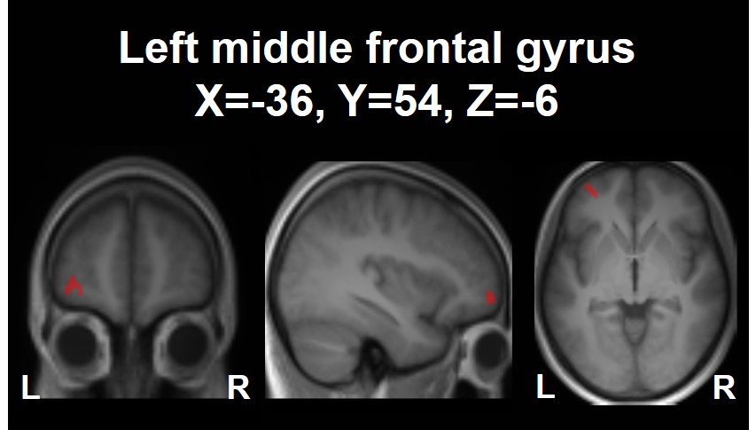
The cluster in the left middle frontal
gyrus (MNI
coordinates: X=-36, Y=54, Z=-6) showing a decrease in μFA upon the 4-week neurocognitive
training. The cluster is shown as an overlay on a 3D-MPRAGE template.
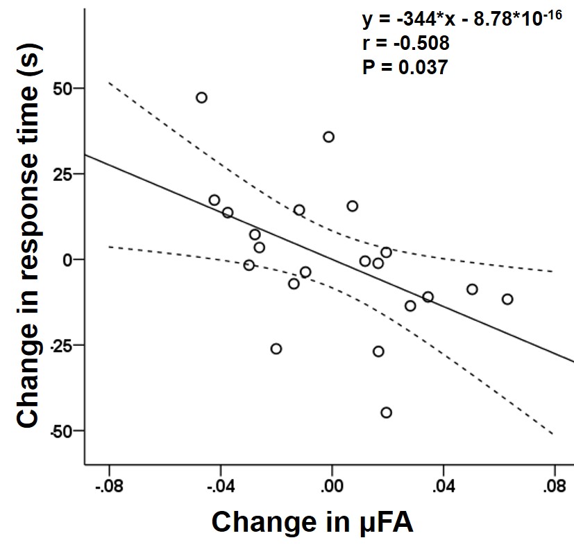
Scatterplots showing significant negative correlation between the change
in μFA of the left middle frontal gyrus and the changes in the response time as
assessed by the orienting attention network test (r=-0.508,
P=0.037). The straight and curved lines indicate the mean and 95% confidence
interval.