Nicolas Delinte1, Claire Gosse2,3, Laurence Dricot3, Quentin Dessain1, Mathieu Simon1, Benoit Macq1, Marie Van Reybroeck2,3, and Gaetan Rensonnet1
1ICTEAM, UCLouvain, Louvain-la-Neuve, Belgium, 2IPSY, UCLouvain, Louvain-la-Neuve, Belgium, 3IoNS, UCLouvain, Brussels, Belgium
1ICTEAM, UCLouvain, Louvain-la-Neuve, Belgium, 2IPSY, UCLouvain, Louvain-la-Neuve, Belgium, 3IoNS, UCLouvain, Brussels, Belgium
This study analyzed multi-shell diffusion MRI on a population of 17 dyslexic children and 18 controls. Advanced models (Diamond & Microstructure Fingerprinting), obtained stronger correlations with children's reading and spelling performance than DTI and showed increased sensitivity.
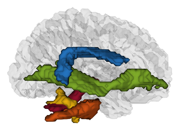
Figure 2. Sagittal view of a 3D representation of the regions of interest described in this report : arcuate fasciculus (in blue), inferior fronto-occipital fasciculus (in green), inferior cerebellar pedunculus (in yellow), middle cerebellar pedunculus (in orange) and superior cerebellar pedunculus (in red).
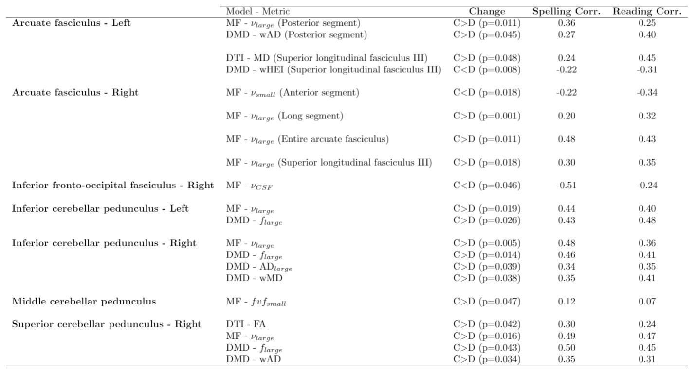
Table 2. Statistically significant differences between the control (C) and dyslexic (D) populations per region and per metric. The models from which the metrics are obtained are mentioned : Diffusion Tensor Imaging (DTI), Microstructure Fingerprinting (MF) or Diamond (DMD). The correlation observed between each metric and the spelling and reading performances in both populations are also provided in the leftmost columns.