Benjamin T Newman1,2, James T Patrie3, and T Jason Druzgal1
1Department of Radiology and Medical Imaging, University of Virginia, Charlottesville, VA, United States, 2Brain Institute, University of Virginia, Charlottesville, VA, United States, 3Department of Public Health Sciences, University of Virginia, Charlottesville, VA, United States
1Department of Radiology and Medical Imaging, University of Virginia, Charlottesville, VA, United States, 2Brain Institute, University of Virginia, Charlottesville, VA, United States, 3Department of Public Health Sciences, University of Virginia, Charlottesville, VA, United States
In the Adolescent Brain Cognitive Development (ABCD) study, we find a positive relationship between an intracellular isotropic diffusion signal and pubertal development in WM regions, providing evidence for complex microstructure changes in brain development within the WM skeleton.
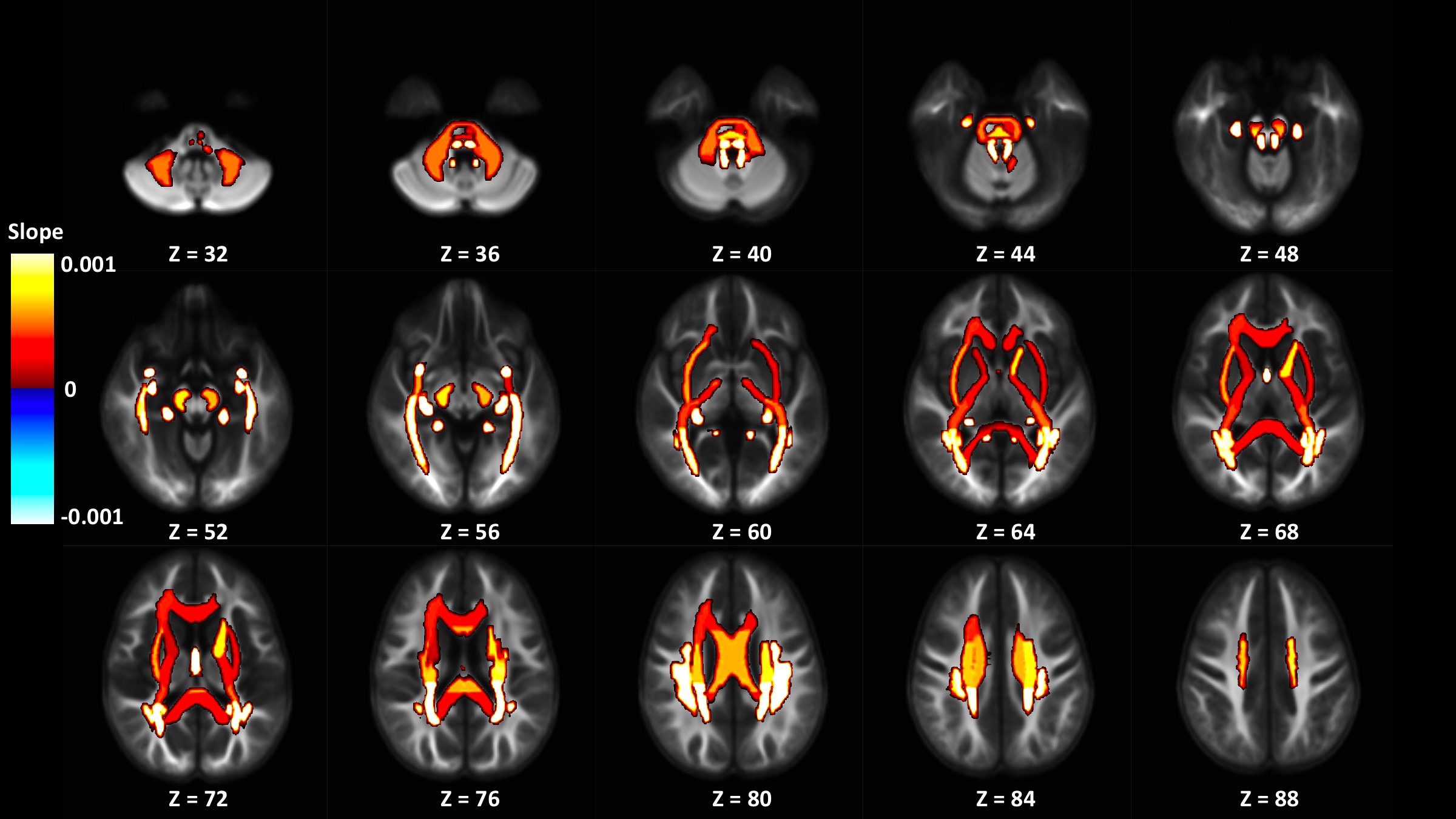
Figure 3: Display of significant adjusted GM-like signal fraction model slopes from ROIs in the JHU WM atlas colored by slope and displayed on the cohort specific template. ROIs located in the posterior parts of the brain appear to be more strongly positively associated with PDSS score than regions elsewhere.
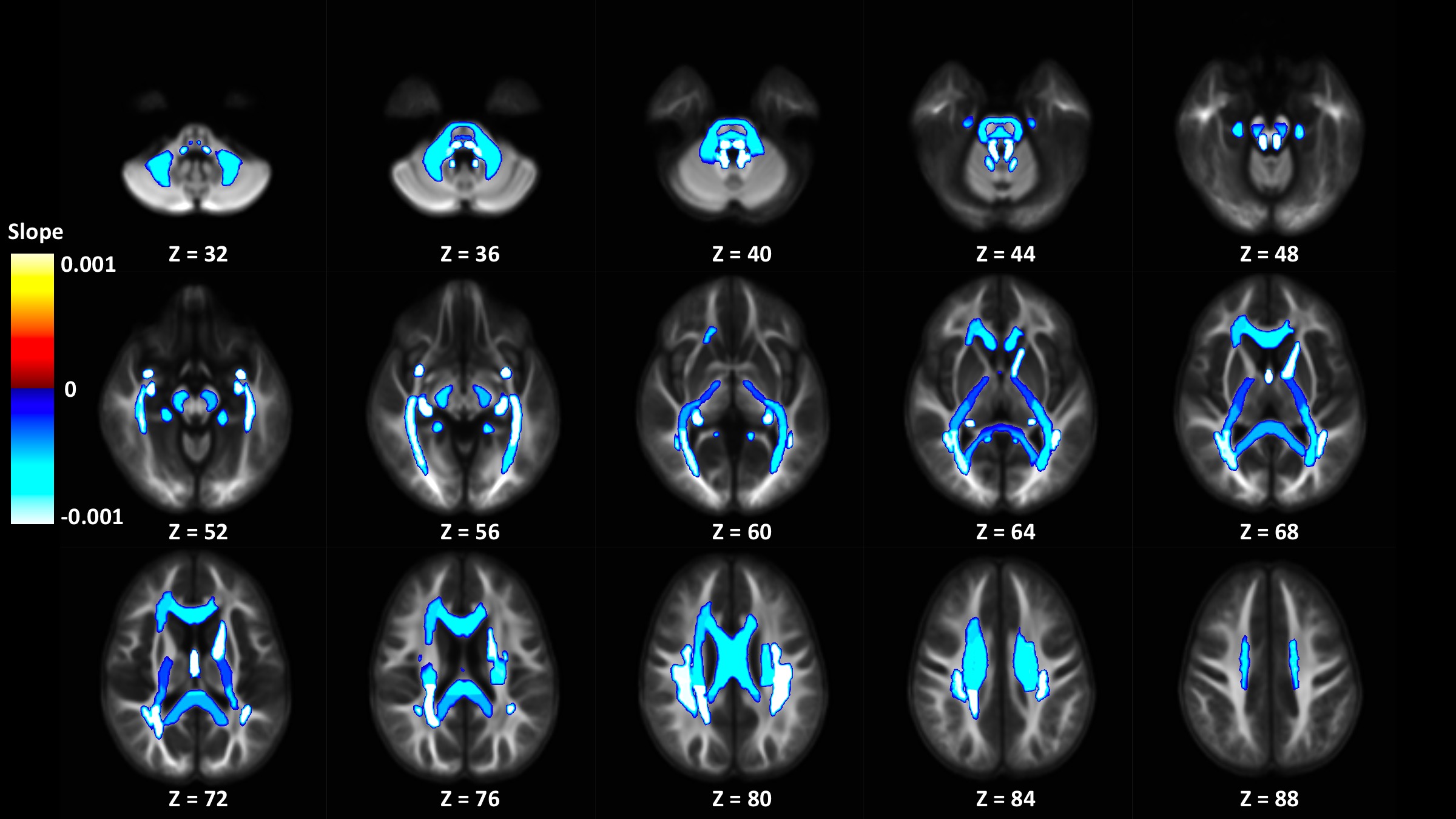
Figure 2: Display of significant adjusted WM-like signal fraction model slopes from ROIs in the JHU WM atlas colored by slope and displayed on the cohort specific template. ROIs located in the posterior parts of the brain appear to be more strongly negatively associated with PDSS score than regions elsewhere.