Loxlan Wesley Kasa1,2, Terry Peters1,2,3,4, Seyed M Mirsattari3,4,5, Ali R Khan1,2,3, and Roy AM Haast1
1Imaging Research Laboratories, Robarts Research Institute, London, ON, Canada, 2School of Biomedical Engineering, Western University, London, ON, Canada, 3Department of Medical Biophysics, Western University, London, ON, Canada, 4Department of Medical Imaging, Western University, London, ON, Canada, 5Department of CNS, Western University, London, ON, Canada
1Imaging Research Laboratories, Robarts Research Institute, London, ON, Canada, 2School of Biomedical Engineering, Western University, London, ON, Canada, 3Department of Medical Biophysics, Western University, London, ON, Canada, 4Department of Medical Imaging, Western University, London, ON, Canada, 5Department of CNS, Western University, London, ON, Canada
Diffusion kurtosis imaging was able to detect possible microstructure anomalies near the temporal pole in both lesional and nonlesional temporal lobe epilepsy subjects, though changes in the nonlesional group were not clearly visible using diffusion tensor imaging metrics.
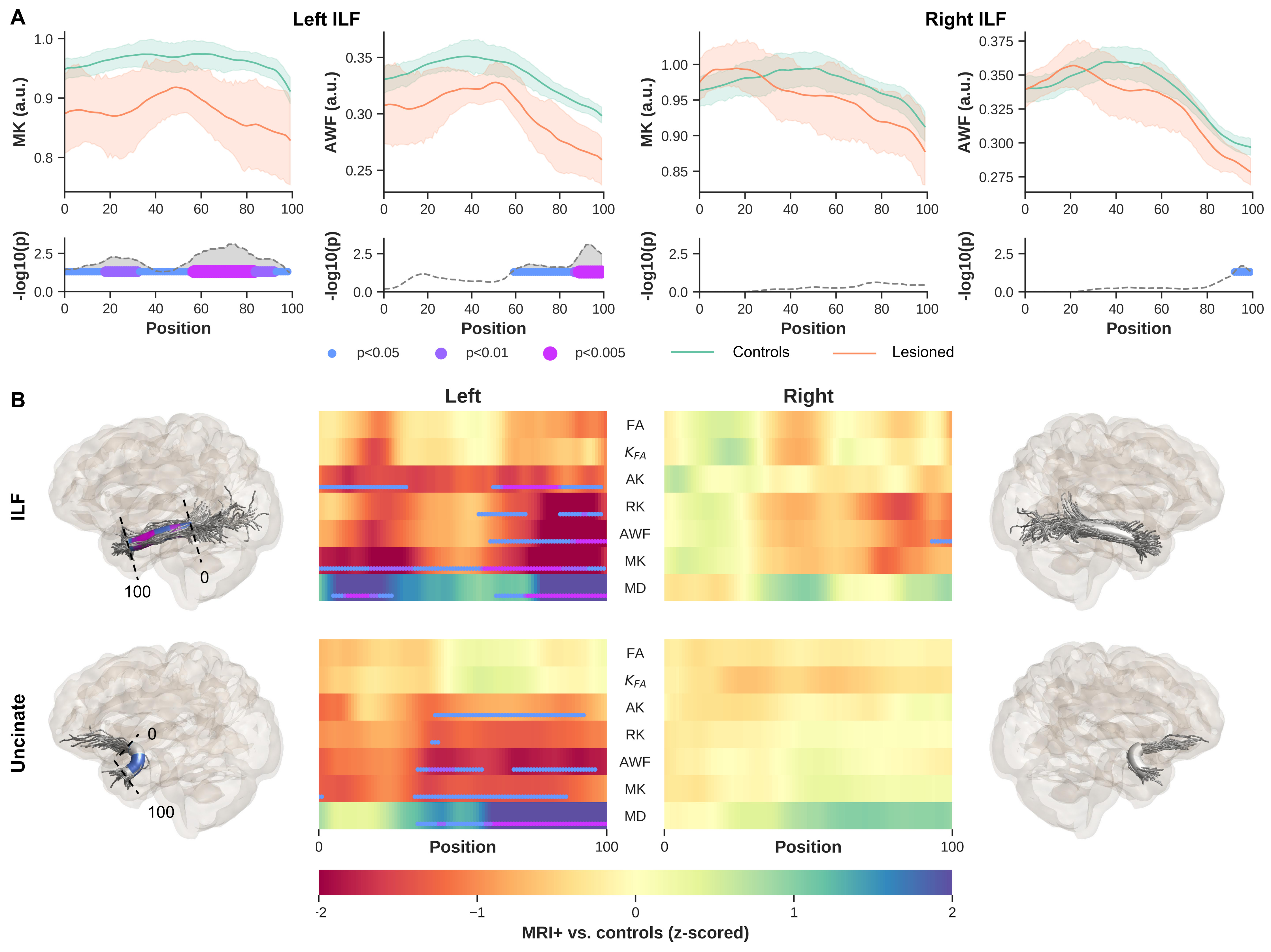
Figures 2. MRI+ patients vs controls. Showing only MK and AWF profiles for left and right ILF (A, top row) and corresponding p-values corrected for multiple comparisons using Bonferroni, age and sex (A, bottom row). Heat maps show z-scores for all DKI derived maps, for ILF (B, top row) and Unc (B, bottom row). To visualize the profile differences at corresponding anatomical locations along the bundles (0-100), the p-values are rendered onto the respective fiber bundles, showing here for MK only. Notice, only results from the side of lesions are shown for each bundle.
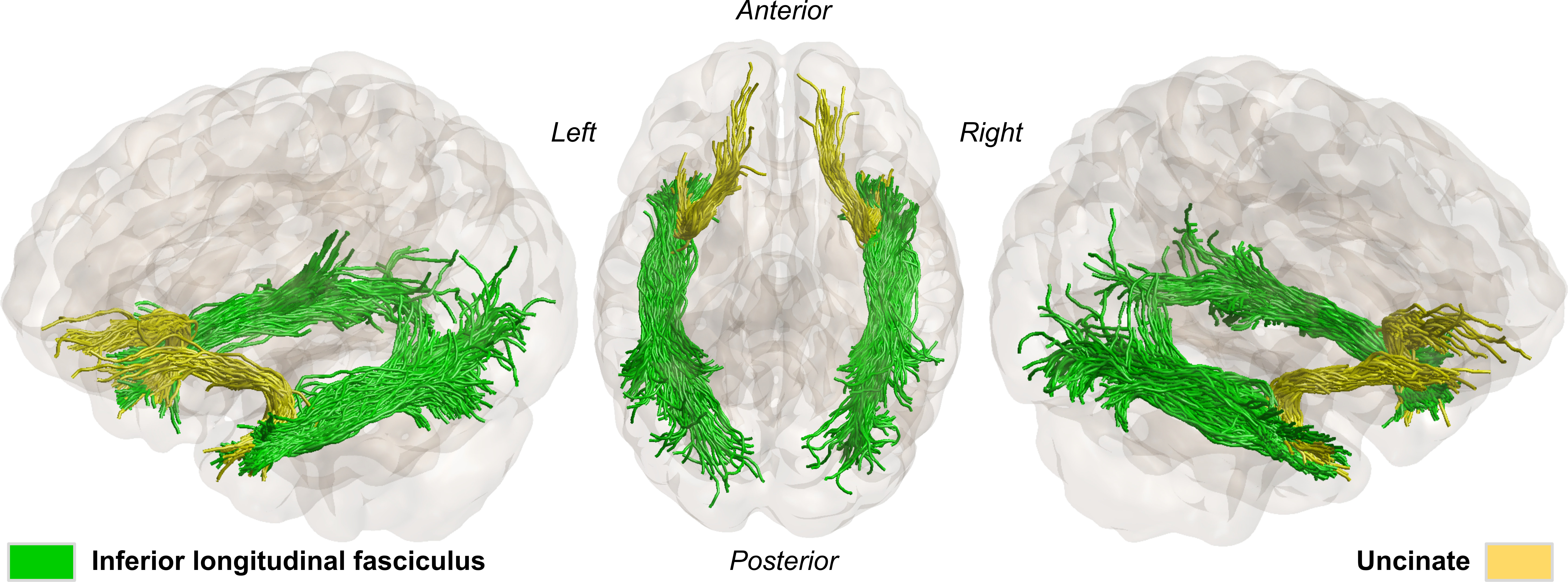
Figures 1. Anatomically constrained tractography, showing the two WM bundles of interest (inferior longitudinal and uncinate fasciculus) for a representative healthy subject.