JIA-WEI Liang1, Tang-Jun Li2, Yao-Wen Liang3, Ting-Chun Lin3, Yi-Chen Lin3, Jiunn-Horng Kang2,4, You-Yin Chen3,5, and Yu-Chun Lo5
1Department of Biomedical Optoelectronic, Taipei Medical University, Taipei, Taiwan, 2College of Medicine, Taipei Medical University, Taipei, Taiwan, 3Department of Biomedical Engineering, National Yang Ming University, Taipei, Taiwan, 4Department of Physical Medicine & Rehabilitation, Taipei Medical University, Taipei, Taiwan, 5Ph.D. Program for Neural Regenerative Medicine, Taipei Medical University, Taipei, Taiwan
1Department of Biomedical Optoelectronic, Taipei Medical University, Taipei, Taiwan, 2College of Medicine, Taipei Medical University, Taipei, Taiwan, 3Department of Biomedical Engineering, National Yang Ming University, Taipei, Taiwan, 4Department of Physical Medicine & Rehabilitation, Taipei Medical University, Taipei, Taiwan, 5Ph.D. Program for Neural Regenerative Medicine, Taipei Medical University, Taipei, Taiwan
We
found that different functional connection between fibromyalgia patients and healthy control participants. The status of
neuroinflammation may play an important role of influencing functional
connection and brain structure may be the part of characteristic feature of fibromyalgia.
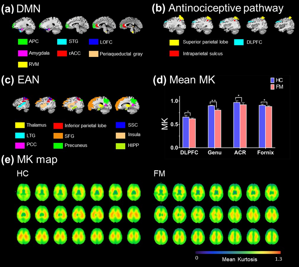
Figure 2. (a) (b) (c) The ROIs were selected form
DMN, antinociceptive pathway and EAN, respectively. (d) Comparison of DKI
parameters showed the significant
differences between HC and FM. In FM, decrease MK values shown in DLPFC
(HC: 0.655 ± 0.00386, FM: 0.617 ± 0.0178 [*p = 0.032]), ACR (control: 0.967 ± 0.036, FM: 0.919 ± 0.042 [**p = 0.007]), fornix (control: 0.907 ±
0.036, FM: 0.88 ± 0.016 [*p
= 0.037]) and genu of corpus callosum (HC: 0.897 ± 0.012, FM: 0.803 ± 0.017 [*p = 0.042]. (e) The MK map of whole
brain shows that decrease MK values.
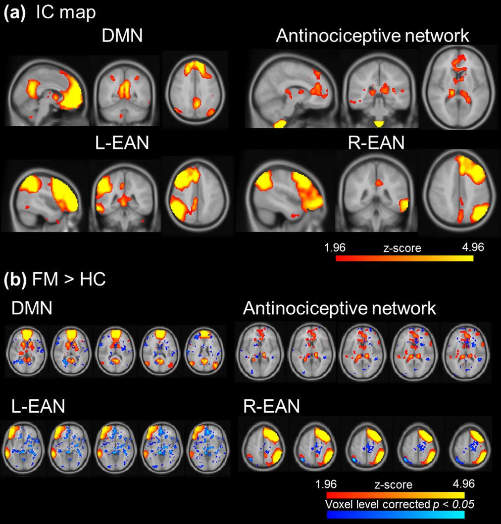
Figure 1. The
visualization of ICA after dual regression. (a) The IC map of DMN, antinociceptive network
and left and right EAN. (b) FM showed significant higher activation
in DMN, antinociceptive network and EAN.