1Department of Radiology, Juntendo University Graduate School of Medicine, Tokyo, Japan, 2Department of Radiological Sciences, Tokyo Metropolitan University, Tokyo, Japan, 3Department of Radiology, Toho University Omori Medical Center, Tokyo, Japan
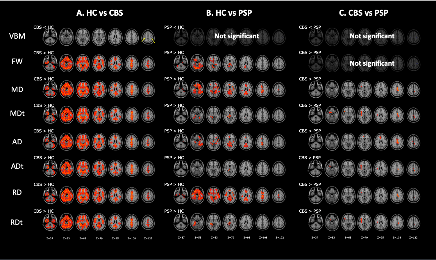
Figure 3. Comparison of VBM, DTI (MD, AD, and RD), and FW imaging (MDt, ADt, RDt, and FW) indices between patients with CBS and PSP and healthy controls.
The results showed reduced gray matter volume (blue–right blue voxels) and increased FW, MD, MDt, AD, ADt, RD, and RDt (red–yellow voxels) in CBS patients relative to controls (A). GBSS results showed increased FW, MD, MDt, AD, ADt, RD, and RDt (red–yellow voxels) in PSP patients relative to controls (B). GBSS results showed increased MD, MDt, AD, ADt, RD, and RDt (red–yellow voxels) in CBS patients relative to PSP patients (C).
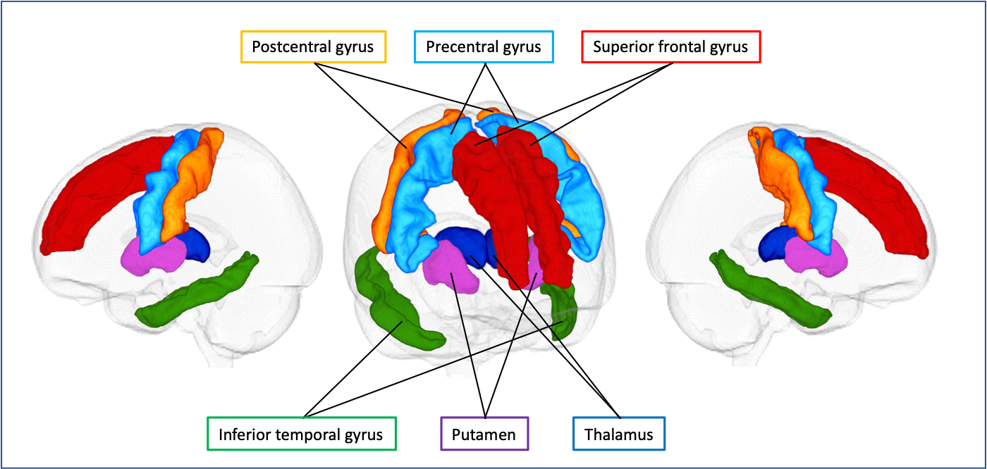
Figure 2. Gray matter regions of interest.
Six gray matter regions, which are described in the previous literature as vulnerable to tau pathology, based on the Desikan–Killiany cortical and subcortical atlas are shown in one representative case.