Xiaoyi He1,2, Weiqiang Dou3, Hansen Schie1, and Junying Wang1,2
1Department of Medical Imaging, Shandong Provincial Qianfishan Hospital, The First Hospital Affiliated with Shandong First Medical University, Jinan, China, 2Shandong First Medical University, Taian, China, 3GE Healthcare, MR Research China, Beijing, China
1Department of Medical Imaging, Shandong Provincial Qianfishan Hospital, The First Hospital Affiliated with Shandong First Medical University, Jinan, China, 2Shandong First Medical University, Taian, China, 3GE Healthcare, MR Research China, Beijing, China
We proved that the combined DKI and 3D-ASL can be effectively tools exploring pathophysiological mechanisms and performing robust diagnostic accuracy for BD patients.
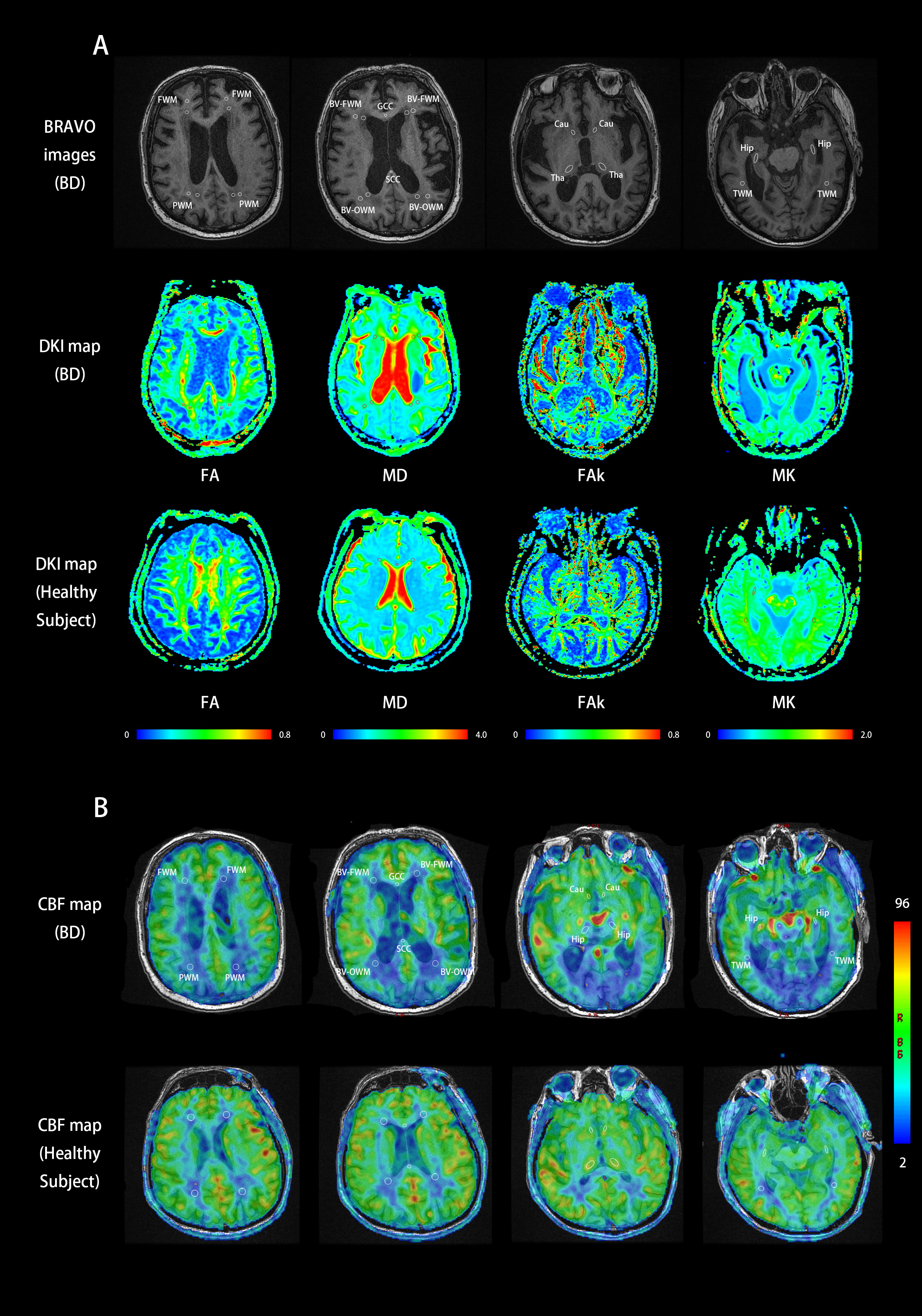
Figure. 1 BRAVO, FA, MD, FAk, MK (A) and CBF (B) maps of BD patients and healthy subjects. 26 kinds of ROIs were drawn along with the area of brain in BD and 18 kinds of ROIs in healthy subjects.
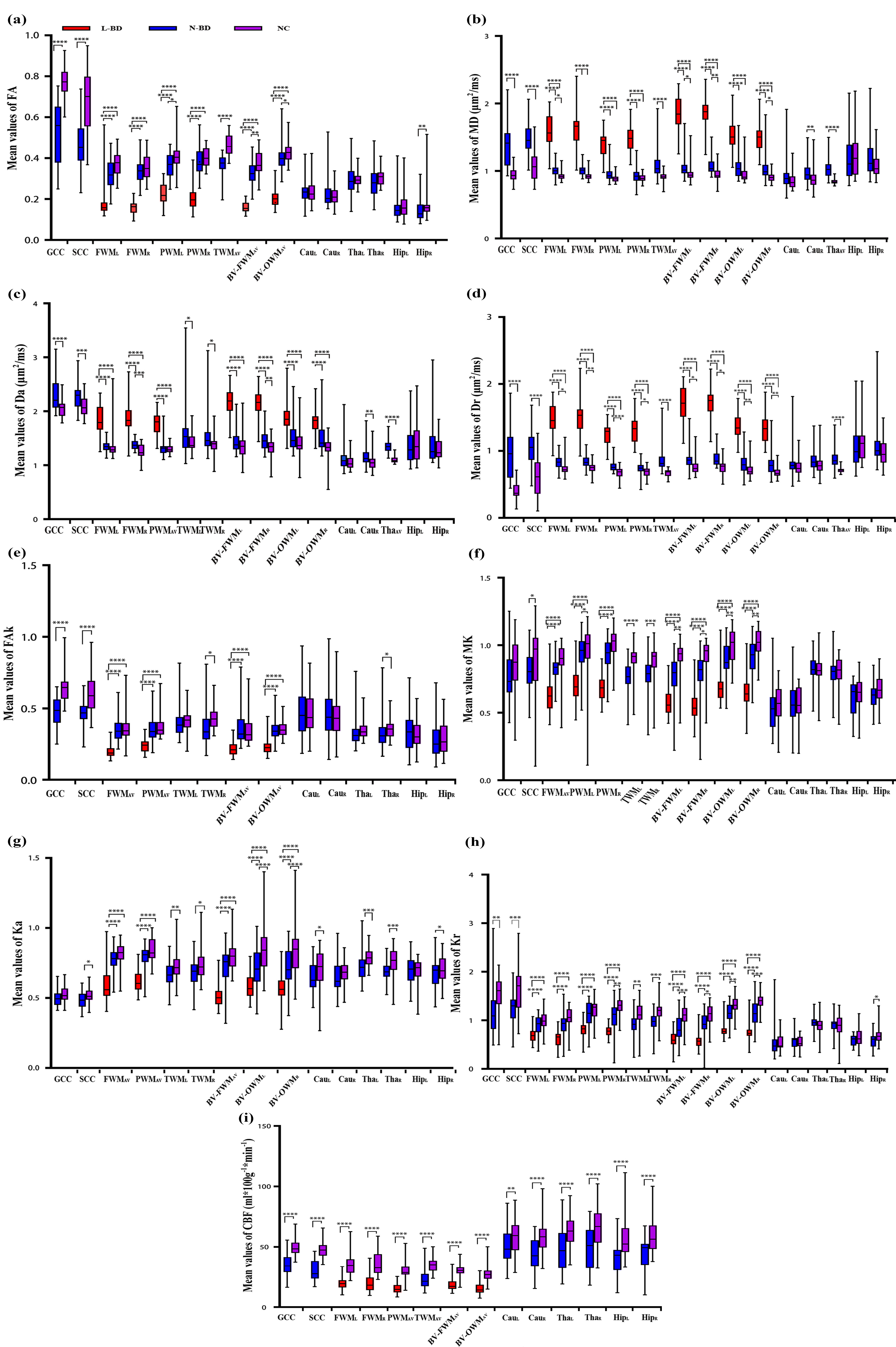
Figure. 2 Comparison of DKI measurements among the three groups and CBF between the different brain regions corresponded in BD and control groups.
Note: L-BD, lesions of BD; N-BD, non-lesions of BD; NC, normal controls; L/R, the left/right-hemispheric side; AV, average of bilateral ROI measurements;GCC/SCC, genu and splenium of the corpus callosum; FWM/PWM/TWM, frontal, parietal, and temporal WM; BV-FWM/BV-OWM, lateral ventricle around frontal and occipital WM; Cau, caudate nucleus; Tha, thalamus; Hip, hippocampus. Significant difference: *p < 0.05; **p <0.001; ***p < 0.0001.