Sisi Li1, Ke Wang2, Xiao Han3, Jinchao Wang3, Wen Jiang3, Xiaodong Ma4, Bing Wu5, Yandong Liu3, Wei Liang3, and Hua Guo1
1Center for Biomedical Imaging Research, Beijing, China, 2Electrical Engineering and Computer Sciences, University of California, Berkeley, Berkeley, CA, United States, 3Beijing Jishuitan Hospital, Beijing, China, 4University of Minnesota, Minnesota, Minnesota, MN, United States, 5GE Healthcare, MR Research China, Beijing, China
1Center for Biomedical Imaging Research, Beijing, China, 2Electrical Engineering and Computer Sciences, University of California, Berkeley, Berkeley, CA, United States, 3Beijing Jishuitan Hospital, Beijing, China, 4University of Minnesota, Minnesota, Minnesota, MN, United States, 5GE Healthcare, MR Research China, Beijing, China
This work investigate the correlation of DTI metrics with the clinical assessment in different WM or GM tracts. The results indicate the potential prognostic value of DTI metrics at non-compressed C2 level for degenerative cervical myelopathy.
Figure
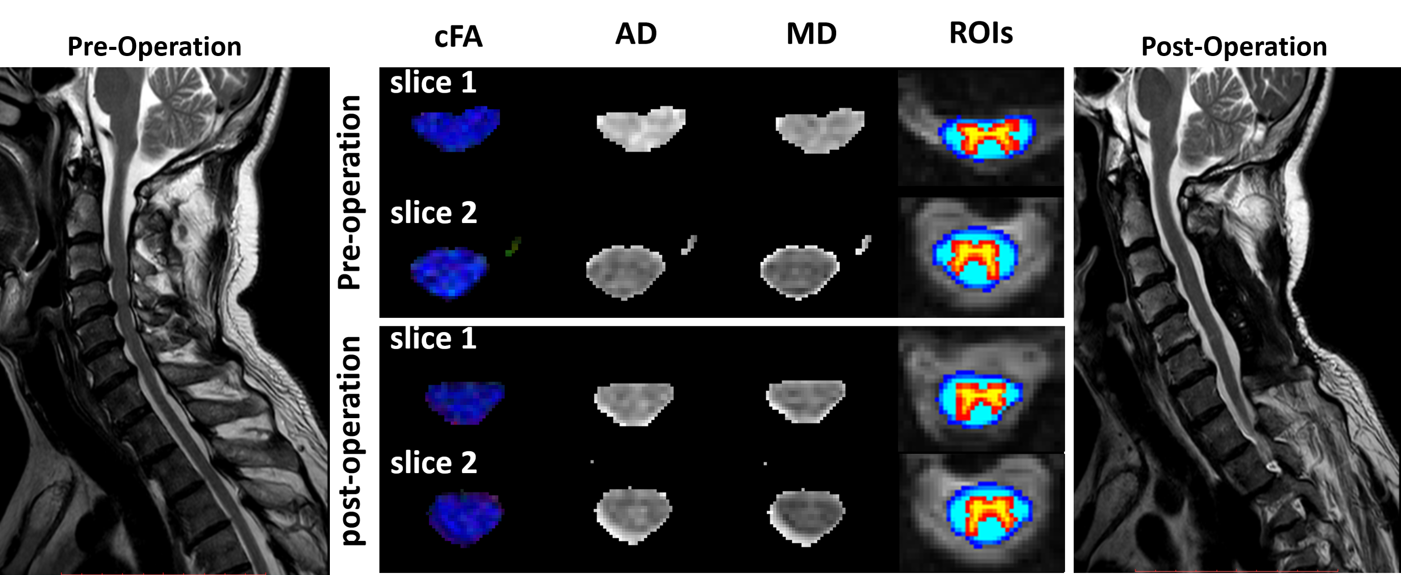
1. Pre-
and post-surgery DTI quantitative maps of one patient with positive surgical
outcome. Slice No.2 represents a vertebral level without visible compression
while slice No.1 represents a compressed level before surgery. WM ROI (blue) and GM ROI (red-yellow) are automatically registered and labeled by SCT.
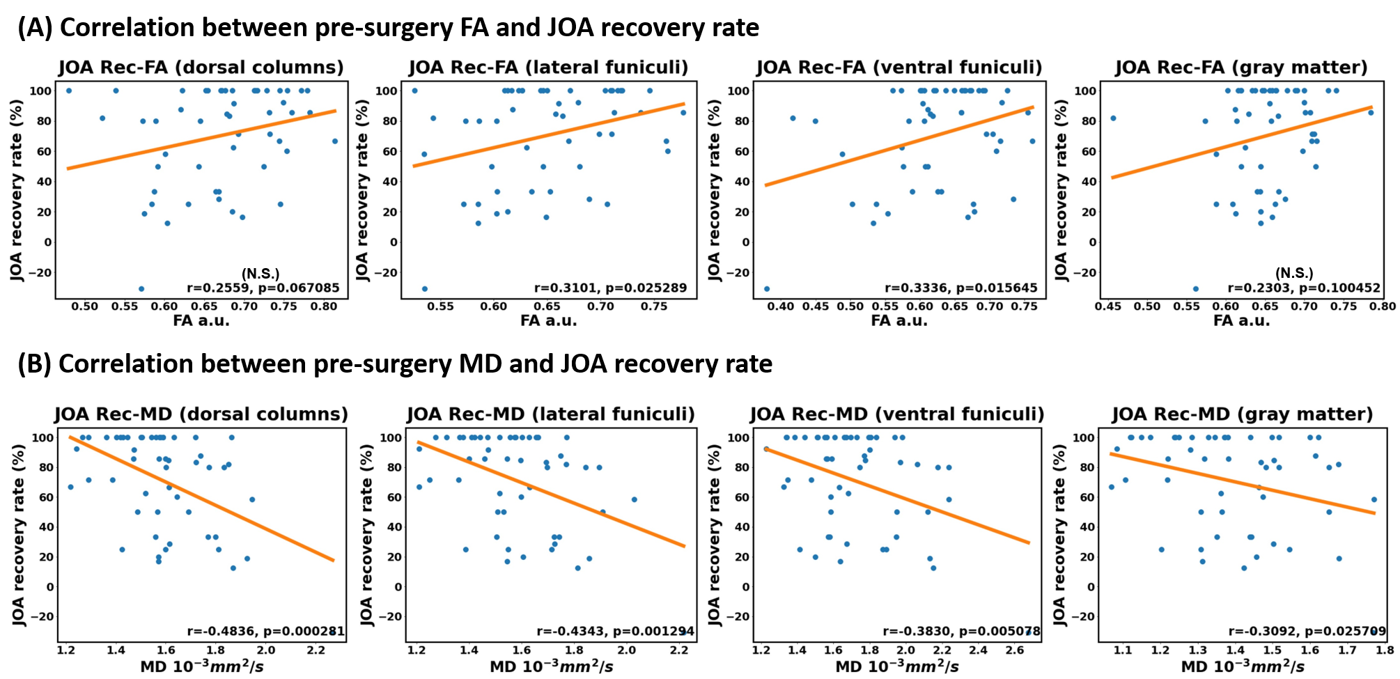
Figure.5
correlation
between pre-surgery (A) FA and (B) MD at C2 level and JOA recovery rate (JOA
Rec) in the following ROIs: dorsal columns (DC), lateral funiculi (LF), ventral
funiculi (VF) and gray matter (GM). The r and P values are marked
in each diagram. Correlation is considered significant if P<0.05.