Cody Johnson1, Ryan Pewowaruk2, David Rutkowski1, Amanda Wolfinger1, and Alejandro Roldán-Alzate1,2,3
1Radiology, University of Wisconsin-Madison, MADISON, WI, United States, 2Biomedical Engineering, University of Wisconsin-Madison, MADISON, WI, United States, 3Mechanical Engineering, University of Wisconsin-Madison, Madison, WI, United States
1Radiology, University of Wisconsin-Madison, MADISON, WI, United States, 2Biomedical Engineering, University of Wisconsin-Madison, MADISON, WI, United States, 3Mechanical Engineering, University of Wisconsin-Madison, Madison, WI, United States
The
sex difference found in LV flow were not found in aortic flow. The VVC of
LV-to-aortic flow was similar for men and women. Dimensional analysis explained
the differences in LV flow as it accounted for differences in cardiac output
and ventricular volume.
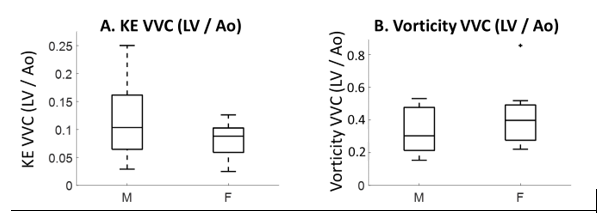
Figure 3: A. Kinetic energy ventricular vascular coupling (VVC), B. vorticity ventricular vascular coupling.
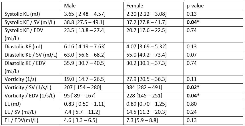
Table 1: Left ventricular flow analysis. where *p<0.05, KE is kinetic energy, mJ is millijoule, SV is stroke volume, EDV is end diastolic volume, L is liter, s is second, EL is energy loss