Yasuhiro Goto1, Michinobu Nagao1, Masami Yoneyama2, Yasutomo Katsumata2, Isao Shiina3, Kazuo Kodaira1, Takumi Ogawa1, Yutaka Hamatani3, Mamoru Takeyama3, Isao Tanaka1, and Shuji Sakai1
1Women's Medical University Hospital, tokyo, Japan, 2Philips Japan, tokyo, Japan, 3Tokyo Women's Medical University Hospital, tokyo, Japan
1Women's Medical University Hospital, tokyo, Japan, 2Philips Japan, tokyo, Japan, 3Tokyo Women's Medical University Hospital, tokyo, Japan
The purpose of this study was to examine mainly LAD about the possibility of the practical use of REPI 4D MRA. REPI 4D MRA with choice of the VENC=30 cm/s could well visualize the coronary arteries from proximal to distal of the LAD.
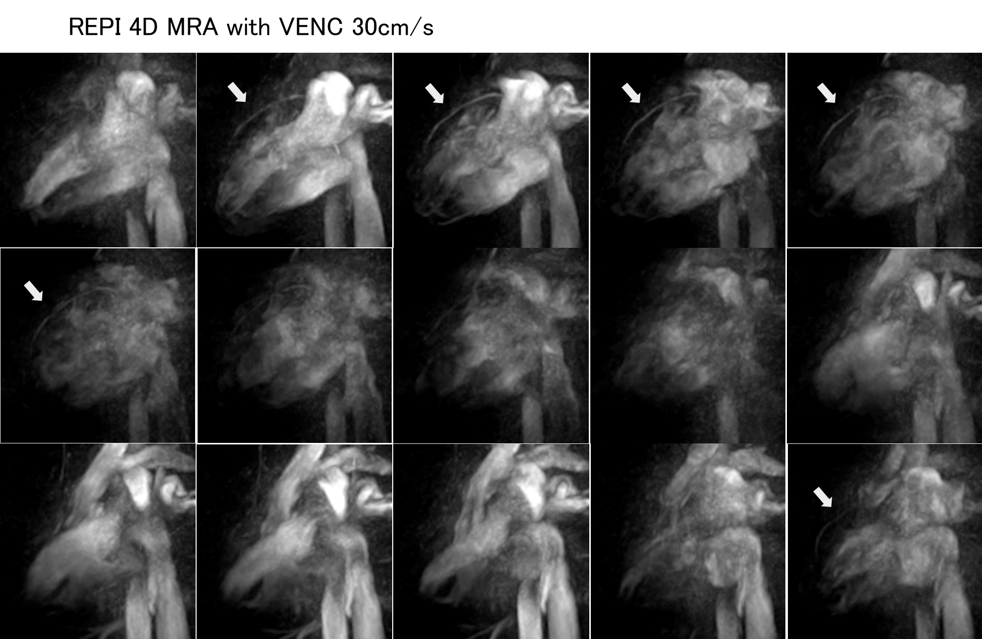
Figure 3. REPI 4d MRA of VENC=30cm/s (upper row).
REPI can be visualized in a well-balanced
manner from the ostial to distal portion of the left
anterior descending artery.
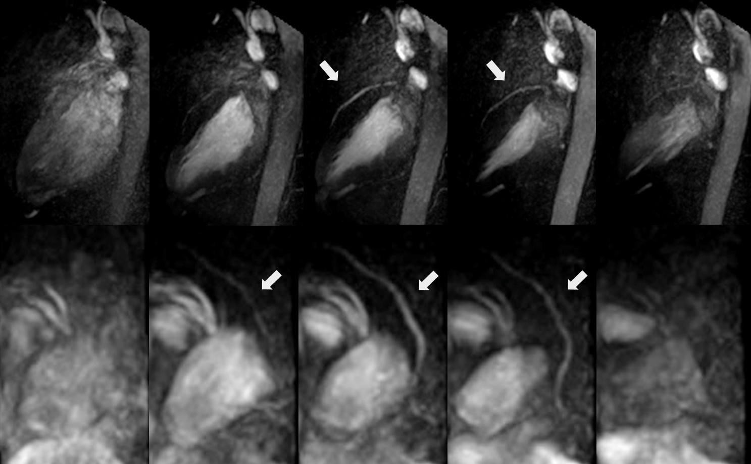
Figure 5. REPI 4D MRA can reconstruct oblique images along the left anterior descending artery (arrow).