Michinobu Nagao1, Yumi Shiina1, Yasuhiro Goto1, Isao Shiina1, Kazuo Kodaira1, Masami Yoneyama2, Takashi Namiki2, Yuka Matsuo1, Atsushi Yamamoto1, Kei Inai1, and Shuji Sakai1
1Tokyo Women's Medical University, Tokyo, Japan, 2Philips Japan, Tokyo, Japan
1Tokyo Women's Medical University, Tokyo, Japan, 2Philips Japan, Tokyo, Japan
Multi-VENC 4D flow MRI with EPI can simultaneously visualize the pulmonary artery stenosis and associated collaterals of slow flow formed by peripheral arteries.
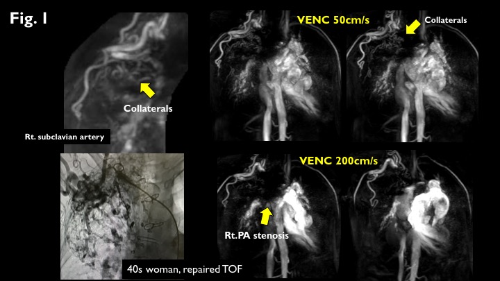
Figure 1.
50s woman with repaired TOF. VENC 50cm/s images can emphasise slow venous flow like collaterals without contrast medium (upper row). Arterial-pulmonary collaterals from the right subclavian artery were detectedas well as angiography (left lower). VENC 200cm/s images show severe stenosis at the proximal right PA (lower row).
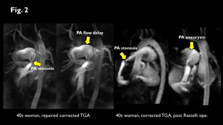
Figure 2.
4D flow with VENC 200cm/s shows typical flow pattern of PA stenosis and aneurysm.