Lihua Chen1, Ailian Liu1, Jiazheng Wang2, Yishi Wang2, and Qingwei Song1
1The First Affiliated Hospital of Dalian Medical University, Dalian, China, 2Philips Healthcare, Beijing, China
1The First Affiliated Hospital of Dalian Medical University, Dalian, China, 2Philips Healthcare, Beijing, China
4D-flow
MRI could measure the increase of flow velocity and volume in both PV and SMV
and a decrease in SV after the meal uptake in healthy volunteers non-invasively.
The APT value of liver parenchyma was found with no change after a meal.
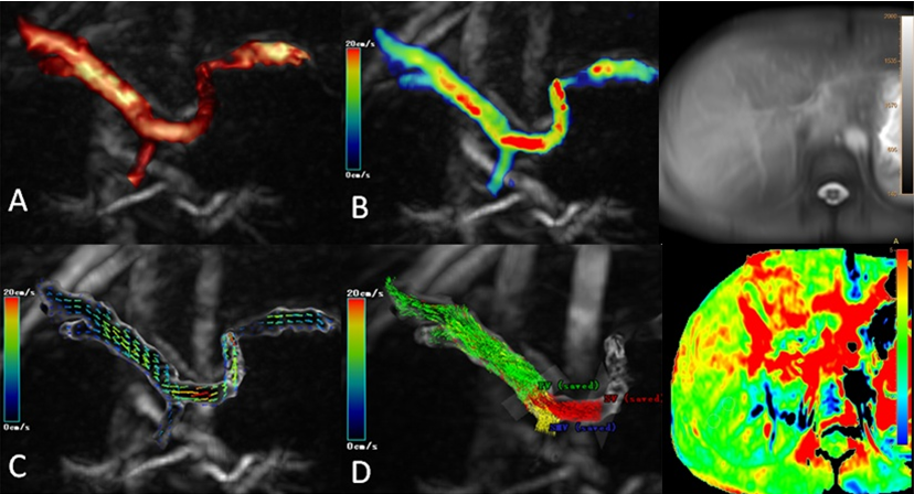
Figure 1 A 22-year-old
female volunteer, reconstructed image (A), flow (B), and direction (C) of portal
system were shown. Red lines representthe SV and yellow lines the SMV
contributions to the total portal blood flow (D), respectively.
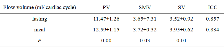
Table
1Comparison of flow volume measurements between fasting and meal