Maurice Pradella1,2, Michael B Scott1, Brad D Allen1, Ryan Avery1, and Michael Markl1,3
1Department of Radiology, Northwestern University, Chicago, IL, United States, 2University Hospital Basel, University of Basel, Basel, Switzerland, 3Department Biomedical Engineering, Northwestern University, Chicago, IL, United States
1Department of Radiology, Northwestern University, Chicago, IL, United States, 2University Hospital Basel, University of Basel, Basel, Switzerland, 3Department Biomedical Engineering, Northwestern University, Chicago, IL, United States
Our full chest, free breathing 4D-flow protocol for clinical
use brought only a minor increase of acquisition time of 3min compared to 2D
phase contrast (2D-PC) in patients requiring aortic and pulmonic evaluation. Respective
flow measurements were consistent in both 4D-flow and 2D-PC.
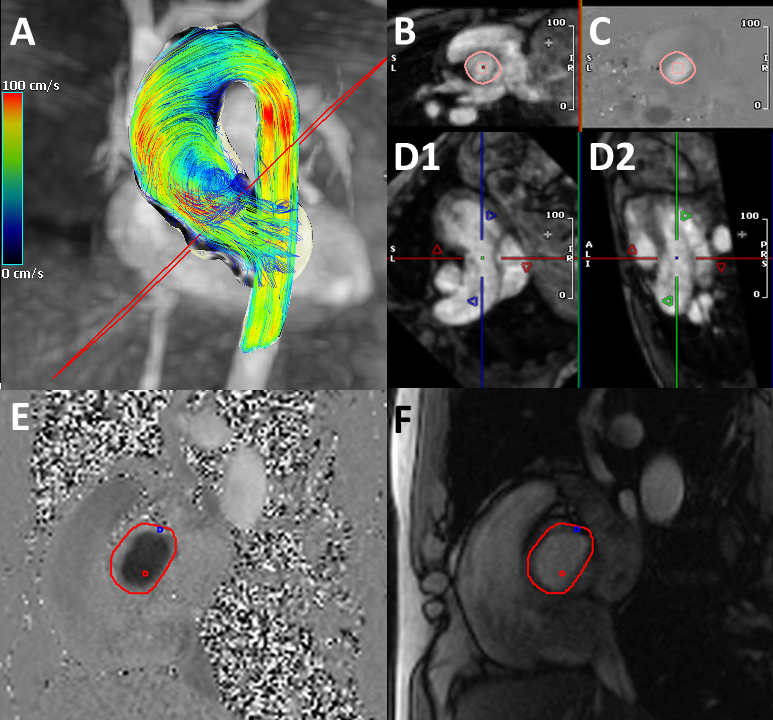
Figure 1: Example of AV measurements. 4D-flow:
A) Color-coded streamline image with corresponding AV measurement plane (red
line) (top left). B) Magnitude and C) Phase contrast images of AV measurement
plane in 4D-flow dataset during systole and D1) & D2) corresponding MPR
images. 2D-PC: E) Phase and F) Magnitude images of AV measurement plane
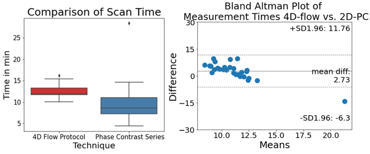
Figure 3: Boxplot (left) and Bland Altman plot
(right) of measurement times between our clinical 4D-flow protocol and the 2D-PC
series. All patients underwent phase contrast series for both aortic and pulmonic
valves.
