Labib Shahid1, James Rice1, Haben Berhane2, Cynthia Rigsby3, Joshua Robinson3, Lindsay Griffin3, Michael Markl2, and Alejandro Roldán-Alzate1
1University of Wisconsin-Madison, Madison, WI, United States, 2Northwestern University, Chicago, IL, United States, 3Ann & Robert H. Lurie Children's Hospital of Chicago, Chicago, IL, United States
1University of Wisconsin-Madison, Madison, WI, United States, 2Northwestern University, Chicago, IL, United States, 3Ann & Robert H. Lurie Children's Hospital of Chicago, Chicago, IL, United States
Computational enhancement of 4D flow MRI improved spatio-temporal resolution, simulated low velocity flow inside aneurysm, and predicted flow inside stent. 4D flow MRI and CFD results agreed. Post-repair analysis showed residual complex flows.
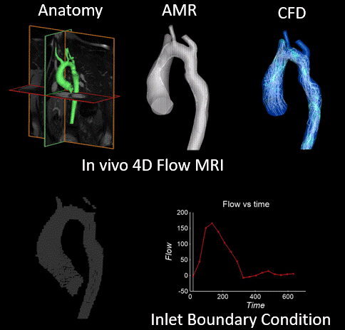
Figure 1: Schematic of computational method enhancing 4D Flow MRI for cardiovascular flow dynamics analysis. Top left: COA patient 2 anatomy pre-repair from CE-MRA. Bottom left: 4D flow MRI study of patient 2 before stent placement. Top center: Autonomously changing computational mesh grid over one cardiac cycle due to AMR. Bottom right: Flow information at ascending aorta extracted from in vivo study used for inlet boundary condition for numerical simulation. Top right: CFD simulation results showing flow information of patient 2 with aortic kinking.
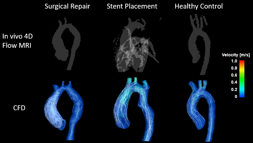
Figure 5: Comparison of in-vivo 4D flow MRI and CFD results for post-interventions cases with healthy control. Good agreement between in-vivo and CFD results in velocity magnitudes at acceleration regions and vortical locations can be seen. Results show residual complex flow regions with larger vortices at ascending aortic section after repair when compared to the healthy case.