Jihye Jang1,2, Yansong Zhao1, Jouke Smink3, Andrew J Powell2, and Mehdi H Moghari2
1Philips Healthcare, Gainesville, FL, United States, 2Department of Pediatrics, Harvard Medical School, Boston, MA, United States, 3Philips Healthcare, Best, Netherlands
1Philips Healthcare, Gainesville, FL, United States, 2Department of Pediatrics, Harvard Medical School, Boston, MA, United States, 3Philips Healthcare, Best, Netherlands
To
improve VNR without velocity aliasing, we developed a novel dual-venc dual-echo 2D cine PC sequence where high and low-venc data are acquired within a single
TR and used for velocity measurement. In 10 patients, the dual-venc PC
demonstrated higher VNR and similar blood flow measurements.
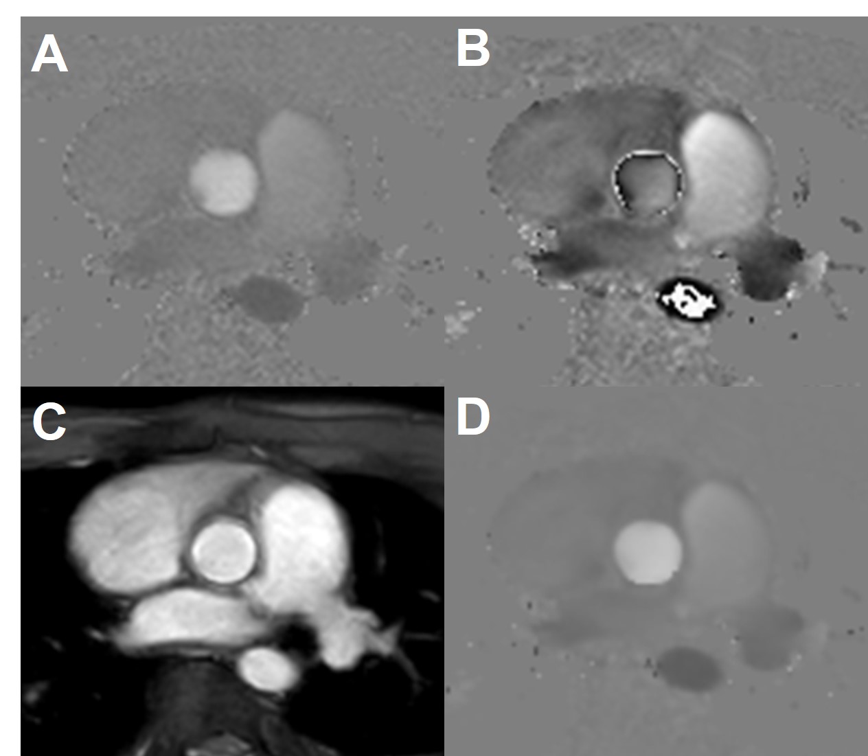
Figure
2. Velocity was measured by unwrapping the velocity of the low-venc and
using the high-venc phase image as an unwrapping threshold. A) Phase image from
a high-venc acquisition with a low VNR. B) Phase image from low-venc
acquisition with a high VNR and velocity aliasing. C) Magnitude image averaged
from both echoes. D) Combined phase image with the VNR of the lower venc image without
the velocity aliasing artifacts.
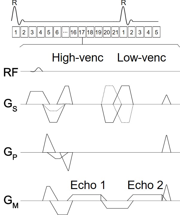
Figure
1. Sequence diagram of a retrospective ECG-gated 2D cine dual-venc
dual-echo PC MRI. High and low-venc data were acquired within a single TR to
minimize the acquisition time associated with the additional venc.