Carmen P S Blanken1, Lukas M Gottwald2, Jos J M Westenberg3, Eva S Peper2, Bram F Coolen4, Gustav J Strijkers4, Aart J Nederveen2, R Nils Planken2, and Pim van Ooij2
1Radiology and Nuclear Medicine, Amsterdam UMC, Amsterdam, Netherlands, 2Radiology and Nuclear Medicine, Amsterdam UMC, location AMC, Amsterdam, Netherlands, 3Radiology, Leiden UMC, Leiden, Netherlands, 4Biomedical Engineering and Physics, Amsterdam UMC, location AMC, Amsterdam, Netherlands
1Radiology and Nuclear Medicine, Amsterdam UMC, Amsterdam, Netherlands, 2Radiology and Nuclear Medicine, Amsterdam UMC, location AMC, Amsterdam, Netherlands, 3Radiology, Leiden UMC, Leiden, Netherlands, 4Biomedical Engineering and Physics, Amsterdam UMC, location AMC, Amsterdam, Netherlands
We show that pseudo-spiral CS 4D flow MRI is at least as reliable as EPI-based 4D flow MRI in measuring blood flow across the heart valves and may be accelerated further to expedite clinical implementation.
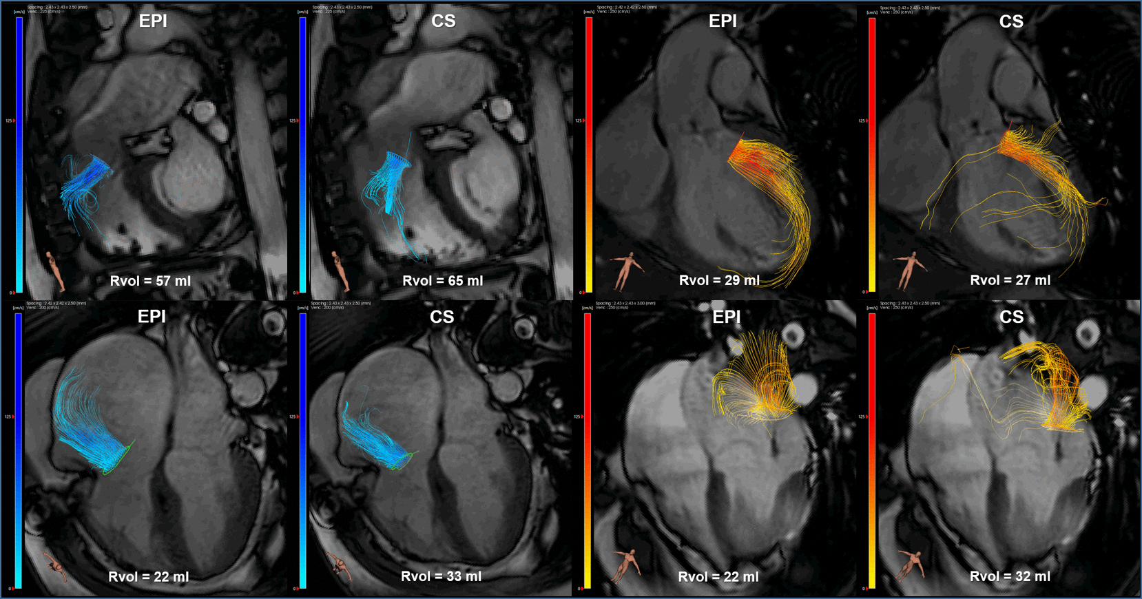
Figure 2: EPI and CS 4D flow MRI streamline visualizations in
four different patients with: pulmonary valve regurgitation (top left), aortic
valve regurgitation (top right), tricuspid valve regurgitation (bottom left)
and mitral valve regurgitation (bottom right). Semi-automated retrospective valve
tracking was performed on bSSFP cine images, on two orthogonal views for each
heart valve. Rvol = regurgitant volume.
Figure
1: EPI and CS 4D flow MRI streamline visualizations of
blood flow through the aortic valve (yellow contour), mitral valve (orange),
pulmonary valve (blue) and tricuspid valve (green) in a 28-year old healthy
volunteer, resulting from semi-automated retrospective valve tracking. Valve
tracking was performed on bSSFP cine images, on two orthogonal views for each
heart valve. 4-chamber bSSFP view is visible in the background.