Eric Schrauben1, Mitzi van Andel2, Lukas Gottwald1, Aart Nederveen1, Maarten Groenink1,2, and Pim van Ooij1
1Department of Radiology & Nuclear Medicine, Amsterdam University Medical Centers, location AMC, Amsterdam, Netherlands, 2Department of Cardiology, Amsterdam University Medical Centers, location AMC, Amsterdam, Netherlands
1Department of Radiology & Nuclear Medicine, Amsterdam University Medical Centers, location AMC, Amsterdam, Netherlands, 2Department of Cardiology, Amsterdam University Medical Centers, location AMC, Amsterdam, Netherlands
This work develops an
open-source 4D flow MRI pulse wave velocity tool. With it, data
sampling requirements are shown to be reducible by an additional 30% to successfully detect differences between
healthy controls and Marfan syndrome patients.
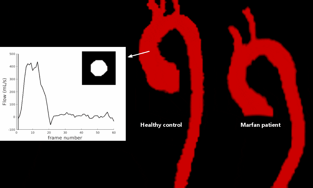
Figure 2. (Animated GIF) Example time-resolved segmentations in subjects
from Figure 1, generated automatically through non-rigid registration of
phase-contrast MR angiographic images at each cardiac time point to a reference
volume. Inset: time-resolved contour in the ascending aorta and resulting flow
waveform over 60 cardiac frames.