Tilman Stephan Emrich1,2,3, Natalie Ring2, U. Joseph Schoepf1, Ning Jin4, Daniel Sebastian Dohle5, Fei Xiong4, Anna Lena Emrich5,6, Karl-Friedrich Kreitner2, and Akos Varga-Szemes1
1Department of Radiology and Radiological Science, Medical University of South Carolina, Charleston, SC, United States, 2Radiology, University Medical Center Mainz, Mainz, Germany, 3DZHK, Partner-Site Rhine-Main, Mainz, Germany, 4Siemens Medical Solutions USA, Inc., Chicago, IL, United States, 5Department of Cardiothoracic Surgery, University Medical Center Mainz, Mainz, Germany, 6Department of Cardiothoracic Surgery, Medical University of South Carolina, Charleston, SC, United States
1Department of Radiology and Radiological Science, Medical University of South Carolina, Charleston, SC, United States, 2Radiology, University Medical Center Mainz, Mainz, Germany, 3DZHK, Partner-Site Rhine-Main, Mainz, Germany, 4Siemens Medical Solutions USA, Inc., Chicago, IL, United States, 5Department of Cardiothoracic Surgery, University Medical Center Mainz, Mainz, Germany, 6Department of Cardiothoracic Surgery, Medical University of South Carolina, Charleston, SC, United States
In our study, both conventional and CS-accelerated 4D
flow techniques provided accurate flow assessment regardless of the presence of contrast
agent, therefore 4D flow
acquisitions can be performed either before or after GBCA administration.
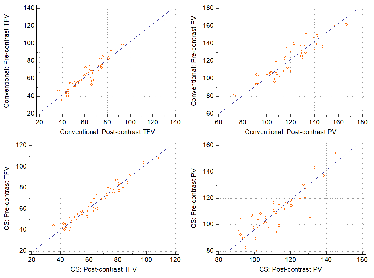
Scatter plots demonstrating correlation between pre-
and post-contrast Total Forward Volume (TFV) and Peak Velocity (PV) measured by
the conventional and CS 4D flow techniques.
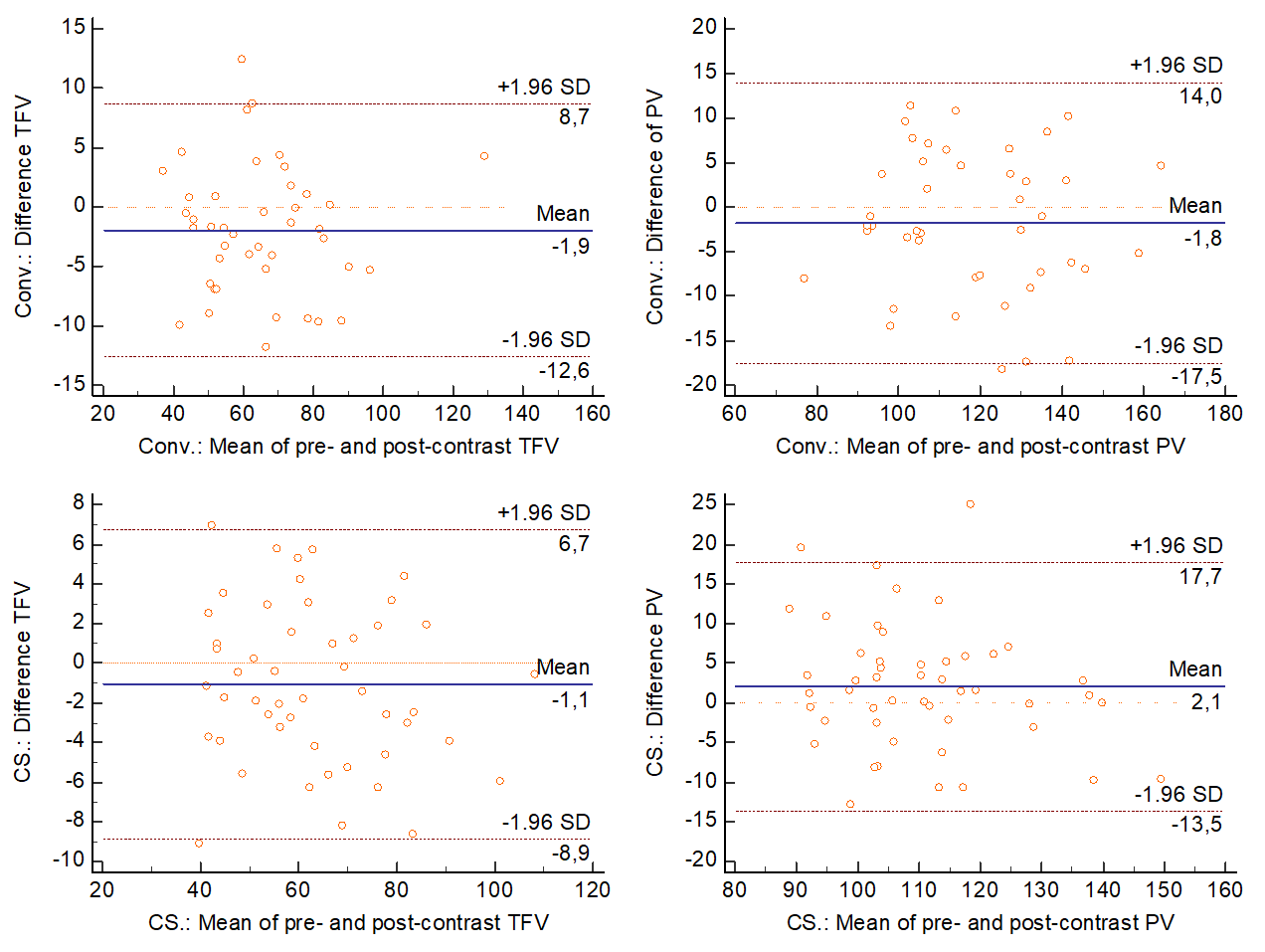
Bland-Altman plots representing the agreement in
pre- and post-contrast Total Forward Volume (TFV) and Peak Velocity (PV)
between the conventional and CS-based 4D flow techniques. Blue solid lines show
the mean of differences, while the dotted lines indicate the upper and lower
limits of agreement (±1.96 SD).