Hajime Tamura1, Hideki Ota2, Tatsuo Nagasaka3, Ryuichi Mori3, Chihiro Kato1, Kohsuke Gonda1, and Kenichi Funamoto4
1Department of Medical Physics, Tohoku University, Graduate school of medicine, Sendai, Japan, 2Department of Advanced MRI Collaboration Research, Tohoku University, Graduate school of medicine, Sendai, Japan, 3Department of Radiology, Tohoku University hospital, Sendai, Japan, 4Institute of Fluid Science, Tohoku University, Sendai, Japan
1Department of Medical Physics, Tohoku University, Graduate school of medicine, Sendai, Japan, 2Department of Advanced MRI Collaboration Research, Tohoku University, Graduate school of medicine, Sendai, Japan, 3Department of Radiology, Tohoku University hospital, Sendai, Japan, 4Institute of Fluid Science, Tohoku University, Sendai, Japan
We designed a 3-dimensional unicursal channel phantom
to simulate small vessels and obtained diffusion-weighted images with varying
infusion rate of water. Comparison of the signal intensities with theoretical simulation
will help understanding the behavior of IVIM imaging.
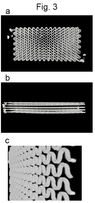
Images obtained by a micro CT unit (SKYSCAN1176, Bruker)
a. A front view. b. A side view. c. A tilted view of one layer.
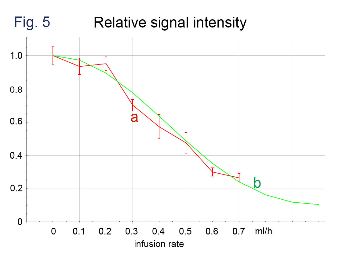
Comparison of (a) signal intensities of phantom
imaging (b = 50 s/mm2, MPG of the phase encoding direction) with (b) those obtained
by computer simulation. The intensities are normalized
to those with the infusion rate = 0.