James Rice1, Labib Shahid1, Haben Berhane2, Joshua Robinson3, Lindsay Griffin3, Cynthia Rigsby3, Michael Markl2, and Alejandro Roldan-Alzate1
1University of Wisconsin-Madison, Madison, WI, United States, 2Northwestern University, Evanston, IL, United States, 3Lurie Children's Hospital, Chicago, IL, United States
1University of Wisconsin-Madison, Madison, WI, United States, 2Northwestern University, Evanston, IL, United States, 3Lurie Children's Hospital, Chicago, IL, United States
In-vitro modeling of COA using 4D flow MRI simulated hemodynamics and improved blood flow visualization of complex flow features. PIV results trended towards those from 4D flow MRI at the center of the aorta, suggesting its use as a method for 4D flow MRI validation of COA hemodynamics.

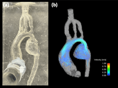
Figure 2: (a) Pre- COA intervention in-vitro silicone model used for flow 4D flow MRI and PIV experiments. (b) Hemodynamic velocity maps obtained by 4D flow MRI with an average inflow rate of 2.6 LPM. Enhanced visualization of complex flow features in the mycotic aneurysm are observed.

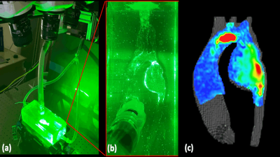
Figure 4: (a) Tomographic (3 camera) PIV setup. A laser is pulsed through a plane cutting a cross section of the model and synced with the acquisition of the high-speed camera. (b) Visualization of particle traces seeded in the aortic flow, illuminated by the laser. The high-speed cameras track the displacement of the particles over the cardiac cycle which are used for velocity quantification. (c) Preliminary velocity map at a cut-plane at the center of the aortic model at peak systole. The high velocity jet extends from the aortic arch to the aneurysmal portion of the descending aorta.