Neeraja Mahalingam1, Andrew Trout2,3,4, Deep Gandhi1, Ruchi Singh5,6, Alexander Miethke5,6, and Jonathan Dillman2,3,5
1Imaging Research Center, Cincinnati Children's Hospital Medical Center, Cincinnati, OH, United States, 2Department of Radiology, Cincinnati Children's Hospital Medical Center, Cincinnati, OH, United States, 3Department of Radiology, University of Cincinnati College of Medicine, Cincinnati, OH, United States, 4Department of Pediatrics, University of Cincinnati College of Medicine, Cincinnati, OH, United States, 5Center for Autoimmune Liver Disease, Cincinnati Children's Hospital Medical Center, Cincinnati, OH, United States, 6Division of Gastroenterology, Hepatology, and Nutrition, Cincinnati Children's Hospital Medical Center, Cincinnati, OH, United States
1Imaging Research Center, Cincinnati Children's Hospital Medical Center, Cincinnati, OH, United States, 2Department of Radiology, Cincinnati Children's Hospital Medical Center, Cincinnati, OH, United States, 3Department of Radiology, University of Cincinnati College of Medicine, Cincinnati, OH, United States, 4Department of Pediatrics, University of Cincinnati College of Medicine, Cincinnati, OH, United States, 5Center for Autoimmune Liver Disease, Cincinnati Children's Hospital Medical Center, Cincinnati, OH, United States, 6Division of Gastroenterology, Hepatology, and Nutrition, Cincinnati Children's Hospital Medical Center, Cincinnati, OH, United States
One-way ANOVA (mixed model) testing showed no significant differences in
ROI-based and whole liver cT1 or MRE-derived liver and spleen stiffness measurements
over a 24 month period in pediatric AILD, although near-significant trends were
observed.
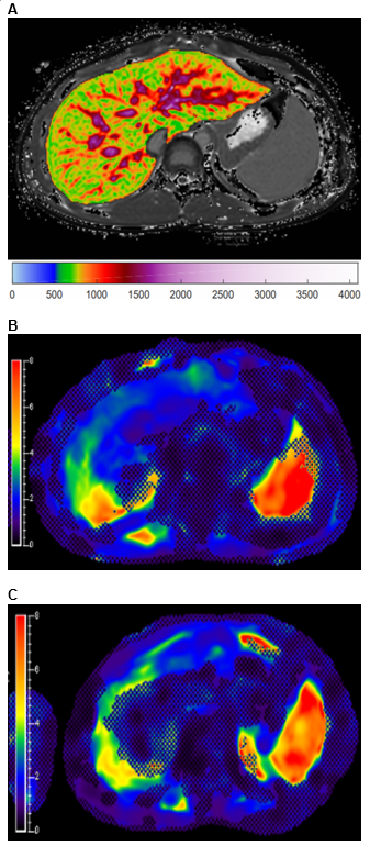
Figure 1: 13-year-old boy with autoimmune sclerosing cholangitis. A) Iron-corrected T1 (cT1) map of the
liver. B) MR elastogram of the liver
(passive driver placed over right upper quadrant). C) MR elastogram of the spleen (passive driver placed over left
flank).
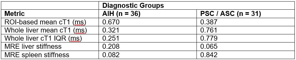
Figure 2: One-way ANOVA (mixed model) results to determine statistical differences
in measurements of interest between baseline, year 1, and year 2 exams for AIH
and PSC / ASC cohorts. P-values reported. AIH: autoimmune hepatitis; PSC:
primary sclerosing cholangitis; ASC: autoimmune sclerosing cholangitis; ROI:
region-of-interest; cT1: iron corrected (T2*) – T1 mapping; MRE:
magnetic resonance elastography.