Xiang-Pan Meng1, Yuan-Cheng Wang1, and Shenghong Ju1
1Radiology, Zhongda Hospital, Medical School of Southeast University, Nanjing, China
1Radiology, Zhongda Hospital, Medical School of Southeast University, Nanjing, China
CT
and MRI had comparable predictive performance for MVI in solitary HCC. Only the radiomics signature at
MRI had significant added value for
MVI prediction on HCC > 2 and ≤ 5 cm.
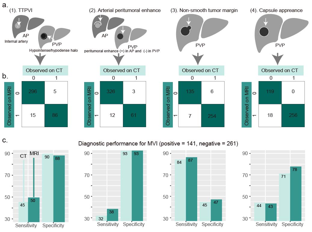
(a) Schematic of two-trait predictor of
venous invasion (TTPVI), arterial peritumoral enhancement, non-smooth tumor
margin, and capsule appearance. (b) Frequencies of presence and absence
of the four features at CT and MRI. The number of cases showing TTPVI, arterial
peritumoral enhancement, non-smooth tumor margin and capsule appearance at MRI
but not at CT was 15, 12, 7, 18, respectively; Number of cases presenting the
four features at CT but not at MRI were 5, 3, 6, and 0, respectively. (c)
Sensitivity and specificity of the four features for MVI diagnosis at CT or
MRI.
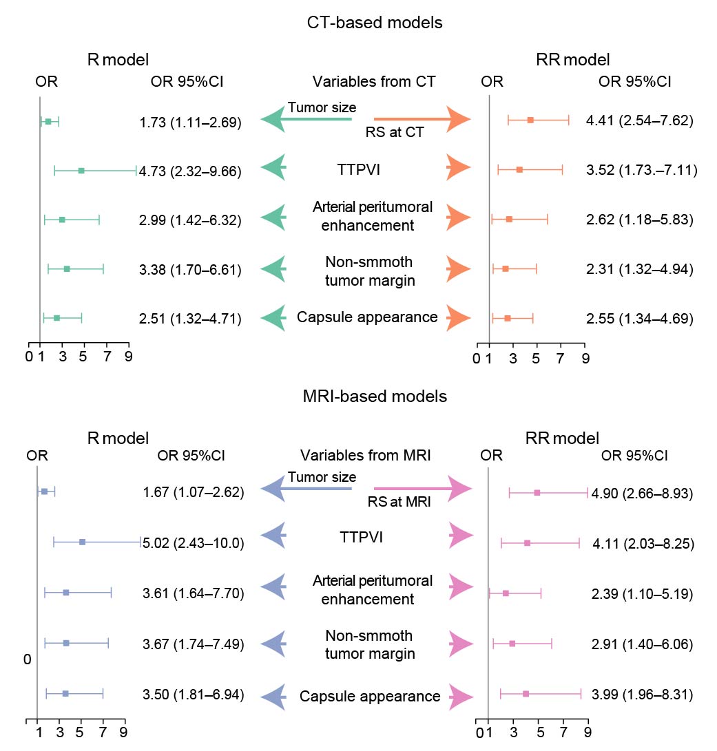
Odds ratios of each
variable contributing to the R and RR models at CT or MRI. R model =
Radiographic model; RR model = radiographic-radiomics model; RS = radiomics
signature; TTPVI = two-trait predictor of venous invasion.