Lili Fan1, Ailian Liu1, Jiazheng Wang2, Liangjie Lin2, Lihua Chen1, Qingwei Song1, Renwang Pu1, Ying Zhao1, Tao Lin1, and Xue Ren1
1The First Affiliated Hospital of Dalian Medical University, Dalian, China, 2Philips Healthcare, Beijing, China
1The First Affiliated Hospital of Dalian Medical University, Dalian, China, 2Philips Healthcare, Beijing, China
Dynamic contrast enhanced imaging (DCE-MRI) combined with T1 and T2 mapping showed a high efficacy to distinguish hepatocellular carcinoma (HCC) from hepatic metastasis (HM) (AUC: 0.936; sensitivity: 81.8%; specificity: 100%).
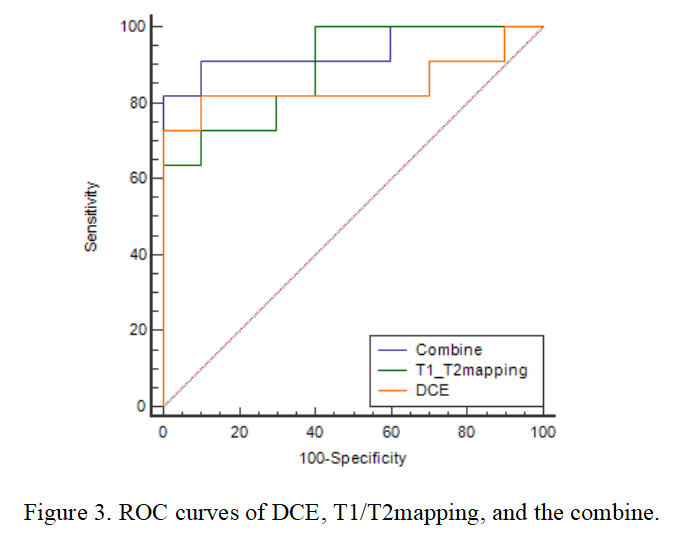
Figure 3. ROC curves of DCE, T1/T2mapping, and the combine.
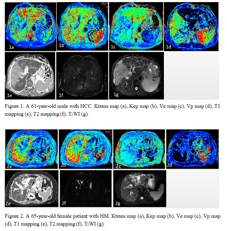
Figure 1. A 61-year-old male with HCC. Ktrans map (a), Kep map (b), Ve map (c), Vp map (d), T1 mapping (e), T2 mapping (f), T2WI (g).
Figure 2. A 65-year-old female patient with HM. Ktrans map (a), Kep map (b), Ve map (c), Vp map (d), T1 mapping (e), T2 mapping (f), T2WI (g).