Bradley C Monteforte1, Ali Agely1, Manoj Mathew1, Pejman Ghanouni1, and Ryan L Brunsing1
1Radiology, Stanford University, Palo Alto, CA, United States
1Radiology, Stanford University, Palo Alto, CA, United States
We found no difference in performance between dedicated 2D-GRE and 3D-SPGR Dixon sequences for the detection of fat or iron content in liver lesions.
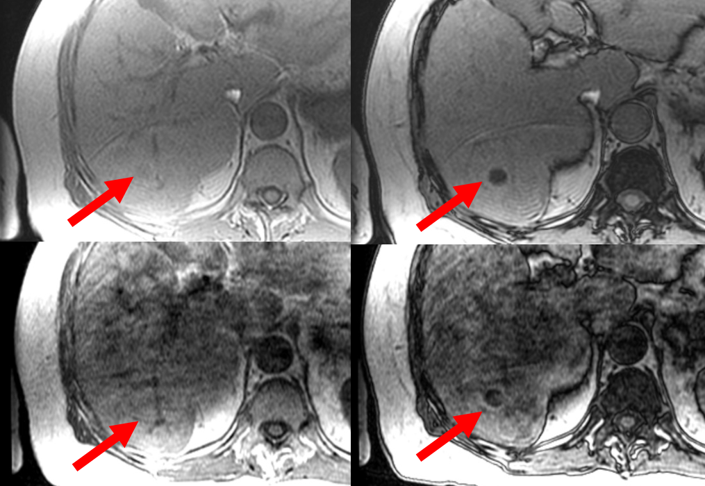
Figure 2. In-phase 2D-GRE
(top left), opposed-phase 2D-GRE (top right), in-phase 3D-SPGR (bottom left),
opposed-phase 3D-SPGR (bottom right). The right hepatic lobe lesion
(arrows) demonstrates signal loss on opposed-phase images with both sequences, as rated
by both readers. Respiratory motion was scored mild and severe by both readers for the 2D-GRE and 3D-SPGR images, respectively. Of note, each pair of IOP
images was assessed at different time points within the full slate of their
respective sequences.
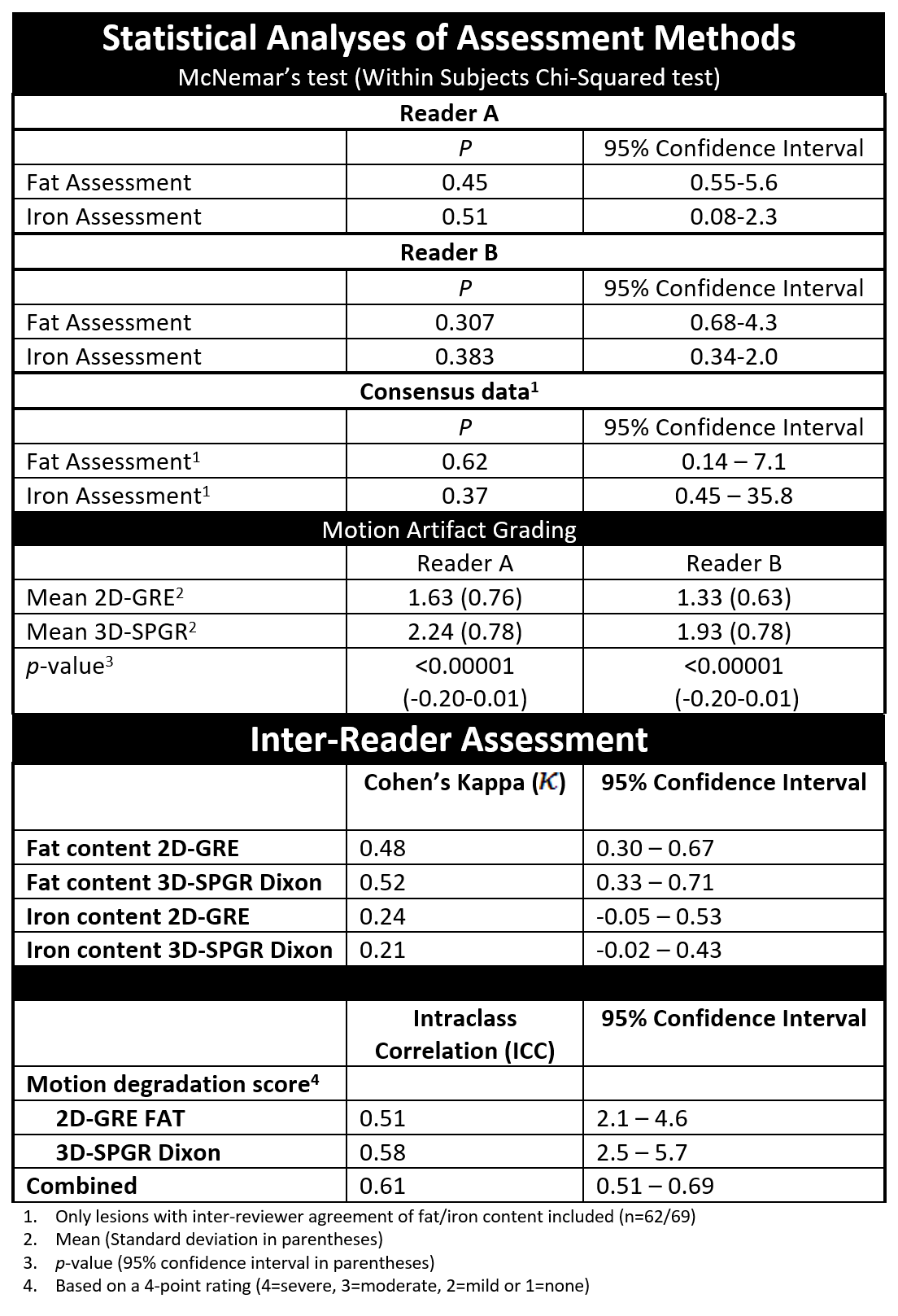
Figure 5. Statistical analyses of assessment methods.