Maike Bode1, Shuo Zhang1,2, Nils A. Krämer1, Christiane K. Kuhl1, and Alexandra Barabasch1
1Diagnostic and Interventional Radiology, University Hospital RWTH Aachen, Aachen, Germany, 2Philips Healthcare, Hamburg, Germany
1Diagnostic and Interventional Radiology, University Hospital RWTH Aachen, Aachen, Germany, 2Philips Healthcare, Hamburg, Germany
Although CS-based reconstruction in
single-shot EPI-DWI can be used to accelerate acquisition time and/or improve
spatial resolution without image quality loss, a small fraction of FLLs went
undetected on CS-DWI.
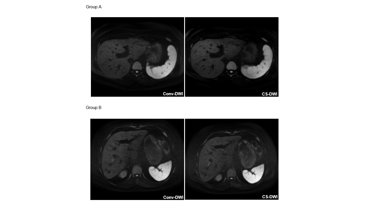
Figure 1. Representative images of conv-DWI and CS-DWI for
group A and group B, which received comparable ratings.
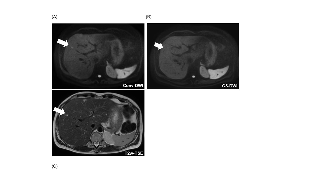
Figure 2. Example of a patient with a missed FLL on CS-DWI.
In conv-DWI (A), a FLL was detected in segment V (arrow), that was not called on CS-DWI (B). In correlation with all sequences from the standard MRI protocol, this FLL corresponded to a thrombosed liver hemangioma.