Anh T. Van1, Sean McTavish1, Johannes M. Peeters2, Kilian Weiss3, Marcus R. Makowski1, Rickmer F. Braren1, and Dimitrios C. Karampinos1
1Department of Diagnostic and Interventional Radiology, School of Medicine, Technical University of Munich, Munich, Germany, 2Philips Healthcare, Best, Netherlands, 3Philips Healthcare, Hamburg, Germany
1Department of Diagnostic and Interventional Radiology, School of Medicine, Technical University of Munich, Munich, Germany, 2Philips Healthcare, Best, Netherlands, 3Philips Healthcare, Hamburg, Germany
When partial Fourier encoding is employed in single-shot diffusion imaging of moving organs phase correction should be performed before the partial Fourier reconstruction to eliminate motion-induced artifacts.
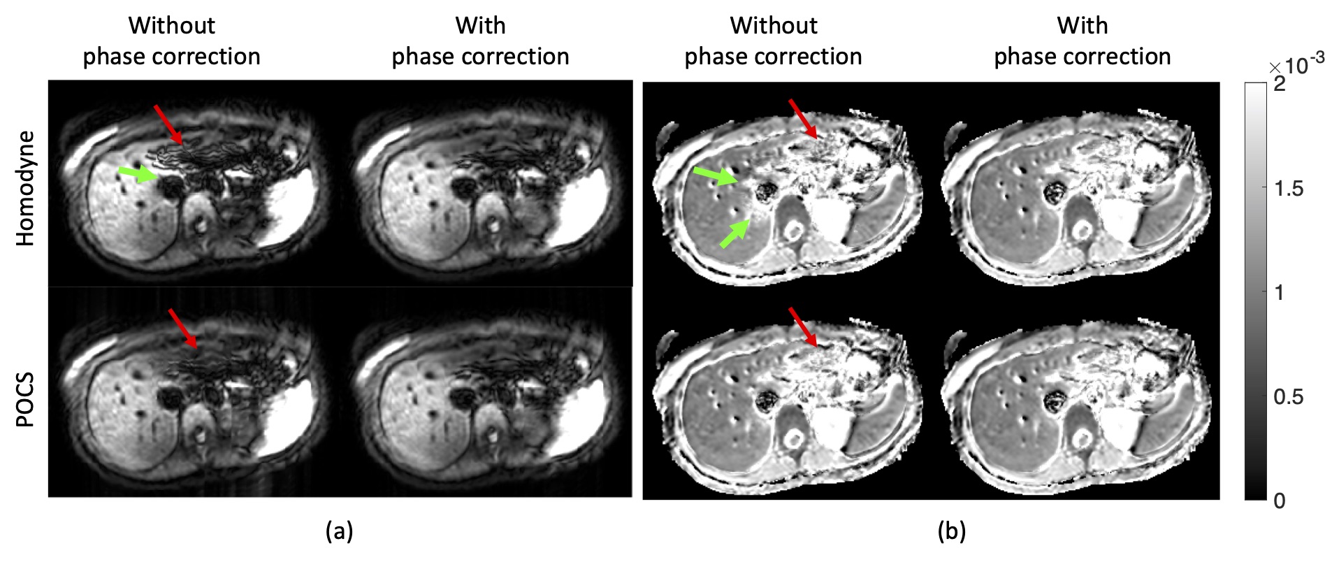
Figure 4. PGSE data with pF = 0.7: single average DWI (a) and ADC maps (b). Without phase correction both homodyne and POCS result in DWI with “worm-like” artifacts in the left liver lobe (red arrows) and with homodyne also around the inferior vena cava (IVC) (green arrows) (a). Phase correction removes the artifact and restore image quality (a). Corresponding ADC maps show an overestimation of ADC around the IVC when homodyne is used without phase correction (b). In the left liver lobe both homodyne and POCS without phase correction yield noisier ADC values than with phase correction.
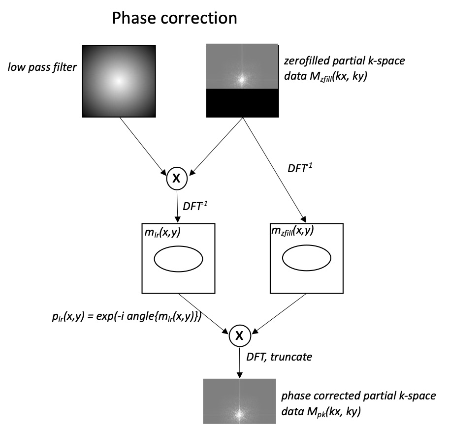
Figure 1. The phase correction procedure before the partial Fourier reconstruction.