Hu Guo1, Huiting Zhang2, Junjiao Hu1, and Jun Liu1
1Department of Radiology, the Second Xiangya Hospital of Central South University, Changsha, China, 2MR Scientific Marketing, Siemens Healthcare, Wuhan, China
1Department of Radiology, the Second Xiangya Hospital of Central South University, Changsha, China, 2MR Scientific Marketing, Siemens Healthcare, Wuhan, China
Compared to SS-EPI, iShim had better image quality and showed more small lesions, and had no significant differences in ADC value.
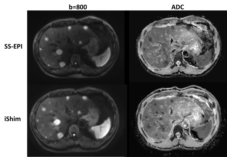
Figure 1. Representative b=800 s/mm2 images and ADC maps using SS-EPI and iShim methods.
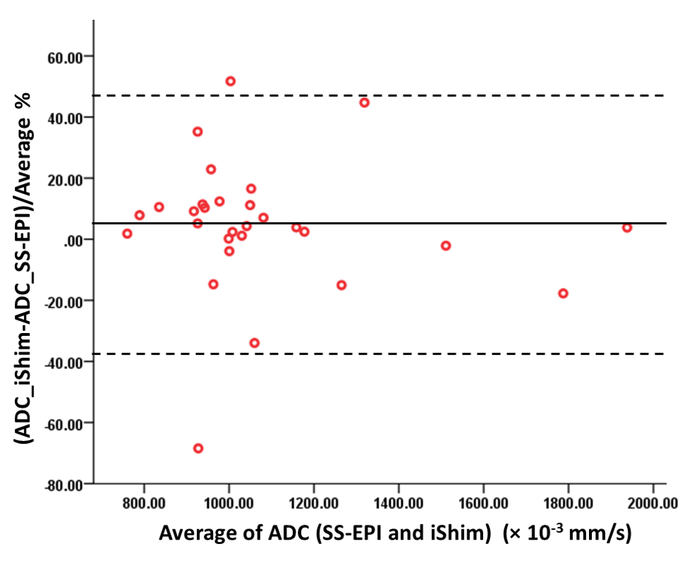
Figure 2. Bland-Altman plot for the ADCs between SS-EPI and iShim sequences. The results indicating the high accordance of ADCs between two sequences.