Nikkita Khattar1, Zhaoyuan Gong1, Matthew Kiely1, Curtis Triebswetter1, Maryam H. Alsameen1, and Mustapha Bouhrara1
1Laboratory of Clinical Investigation, National Institute on Aging, Baltimore, MD, United States
1Laboratory of Clinical Investigation, National Institute on Aging, Baltimore, MD, United States
An
artificial neural network model was trained and successfully used to generate
myelin water fraction maps from conventional relaxation times and proton
density maps.
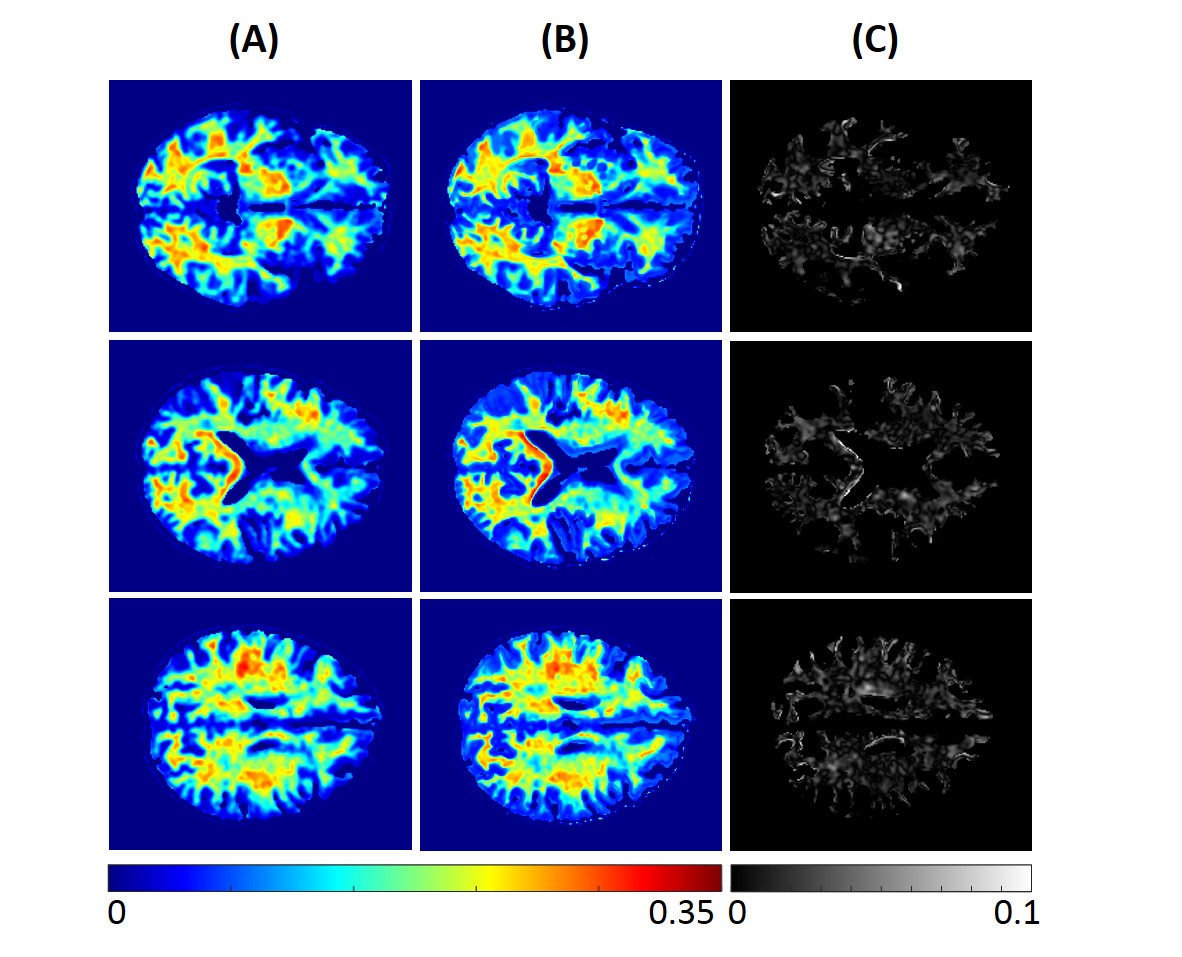
Figure 1. MWF maps from the brain imaging of a young
participant. Results are displayed for three different axial slices. (A)
represents the MWF maps calculated from BMC-mcDESPOT method (the reference
method). (B) represents MWF maps calculated using our trained neural network
(NN) model. (C) shows the absolute difference map between the reference and the
NN methods.
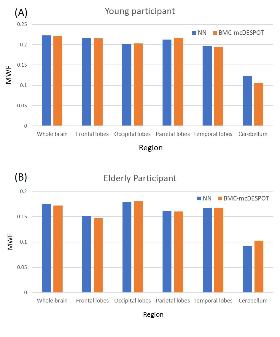
Figure 3. Mean MWF values calculated within representative
white matter brain regions using the NN (blue) and BMC-mcDESPOT (orange)
methods. Results are shown for both the young (A) and elderly (B) participants,
and indicate that NN- and BMC-mcDESPOT-derived MWF exhibit virtually similar
values for all regions evaluated.