Camila Munoz1, Sam Ellis1, Stephan G Nekolla2, Karl P Kunze1,3, Teresa Vitadello4, Radhouene Neji1,3, René M. Botnar1, Julia A. Schnabel1, Andrew J. Reader1, and Claudia Prieto1
1School of Biomedical Engineering and Imaging Sciences, King's College London, London, United Kingdom, 2Nuklearmedizinische Klinik und Poliklinik, Technische Universitat Munchen, Munich, Germany, 3MR Research Collaborations, Siemens Healthcare Limited, Frimley, United Kingdom, 4Department of Internal Medicine I, University hospital rechts der Isar, Technical University of Munich, Munich, Germany
1School of Biomedical Engineering and Imaging Sciences, King's College London, London, United Kingdom, 2Nuklearmedizinische Klinik und Poliklinik, Technische Universitat Munchen, Munich, Germany, 3MR Research Collaborations, Siemens Healthcare Limited, Frimley, United Kingdom, 4Department of Internal Medicine I, University hospital rechts der Isar, Technical University of Munich, Munich, Germany
We introduce a cardiac PET-MR image reconstruction framework that uses high quality diagnostic MR images and MR-derived motion fields to enable PET motion correction and anatomical guidance, resulting in sharper, noise-suppressed cardiac PET images.
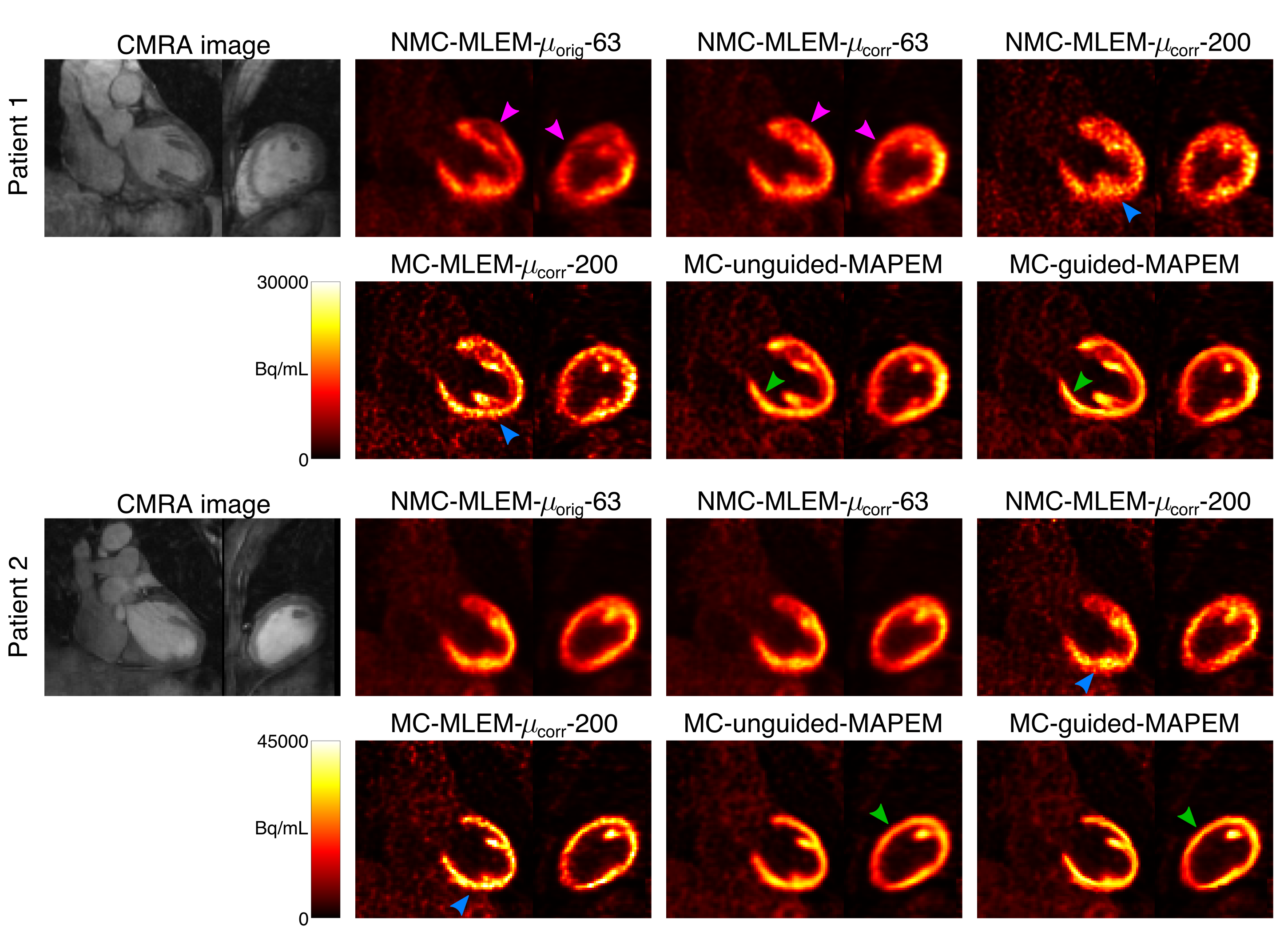
Fig 2. Comparative reconstruction methods for two oncology patients alongside corresponding CMRA images. Aligning the scanner-provided μ-map to the CMRA image removes a defect mimicking artefact in P1 (magenta arrows), while applying motion compensation improves contrast and sharpness in the inferior myocardium (blue arrows). Using either unguided or MR-guided regularization (β=400) reduces noise, however MR-guidance results in better edge-preservation and contrast (green arrows).
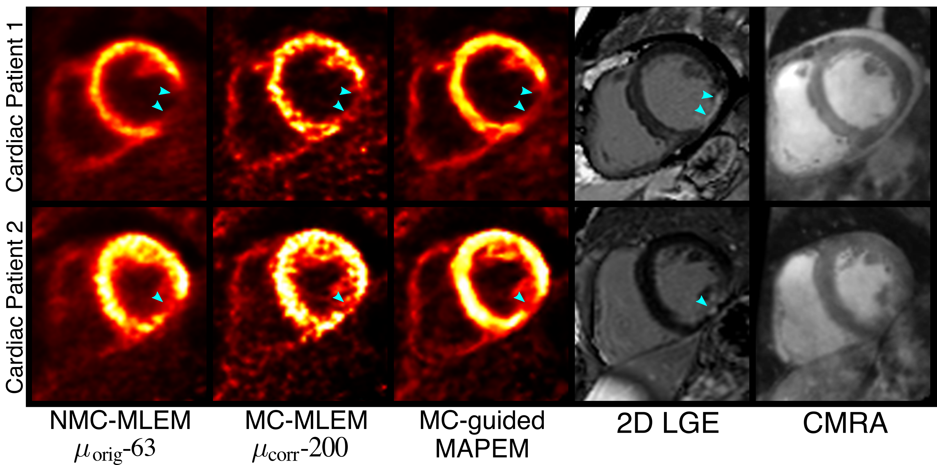
Fig 4. Example short-axis views of the reconstructed PET images for two cardiac patients, and corresponding 2D LGE images showing the extent of myocardial scarring. Using the proposed MC-guided MAPEM (with automatic β selection) method improves image quality while maintaining the appearance of myocardial defects (cyan arrows). Note that LGE images are shown only for comparison and did not provide any information for the MR-guided PET reconstructions, which instead used high resolution CMRA images (also shown here).